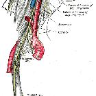
Arteria cerebelli inferior posterior (PICA)

Posterior
inferior cerebellar artery • Posterior inferior cerebellar artery (PICA) infarct - Ganzer Fall bei Radiopaedia

The brain and
arteries at base of the brain. Circle of Willis is formed near center. The temporal pole of the cerebrum and a portion of the cerebellar hemisphere have been removed on the right side. Inferior aspect (viewed from below).


Posterior
inferior cerebellar artery • PICA infarct - Ganzer Fall bei Radiopaedia

Posterior
inferior cerebellar artery • Fourth ventricle posterior inferior cerebellar artery aneurysm - Ganzer Fall bei Radiopaedia

Posterior
inferior cerebellar artery • PICA - caudal loop and choroid point (annotated image) - Ganzer Fall bei Radiopaedia

Posterior
inferior cerebellar artery • Absent right PICA - Ganzer Fall bei Radiopaedia

Anterior
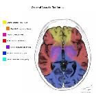
inferior cerebellar artery • Posterior fossa vascular territories (illustration) - Ganzer Fall bei Radiopaedia

Anterior
inferior cerebellar artery • Cerebellar arteries (annotated image) - Ganzer Fall bei Radiopaedia

Anterior
cerebral artery • Common variants of the circle of Willis (illustrations) - Ganzer Fall bei Radiopaedia

Anterior
inferior cerebellar artery • Brainstem arterial territories (diagrams) - Ganzer Fall bei Radiopaedia
Posterior inferior cerebellar artery (PICA) is one of the three vessels that provide arterial supply to the cerebellum. It is the most variable and tortuous cerebellar artery.
Gross anatomy
Origin
Its origin is highly variable:
- ~20% arise extracranially, inferior to the foramen magnum
- 10% arise from the basilar rather than vertebral artery
- 2% bilaterally absent
- occasionally loops around the cerebellar tonsil
Segments
- curves forming the 'caudal loop' which is located anteroinferior to the tip of the cerebellar tonsil, but does not relate to the tonsillar position
- the apex of the caudal loop is
- above foramen magnum in 60% of cases
- at the level of foramen magnum in 10%
- and below the foramen in 30%
- its relationship to the vertebral artery is also variable
- 84% lateral
- 16% medial
- ascends posterior to the medulla behind CN IX and CN X and along the inferior medullary velum
- junction between the posterior medullary segment and the supratonsillar segment is upwardly convex and is the site of origin of small choroidal branches
- known as the choroid point, choroid arch or cranial loop
- this point has a constant relationship with the 4th ventricle and was used prior to cross-sectional imaging to assess for a shift in its position
Branches
- anterior and lateral medullary segments
- small perforating medullary branches (absent in 50%)
- supratonsillar segment
- tonsillohemispheric (lateral) branch
- inferior vermian (median) branch
Note: occasionally, a small vertebral will terminate into a common PICA/AICA trunk.
Supply
It has a variable territory depending on the size of the AICA (AICA-PICA dominance). Typically it supplies:
- posteroinferior cerebellar hemispheres (up to the great horizontal fissure)
- cerebellar tonsils: 85% of the time
- biventral lobule: 80%
- nucleus gracilis: 85%
- superior semilunar lobule: 50%
- inferior portion of the vermis
- lower part of the medulla: 50%
- inferior cerebellar peduncles
See also
Siehe auch:
- Cerebellum
- Arteria cerebelli superior
- posterior inferior cerebellar artery (PICA) infarct
- Nervus glossopharyngeus
- Arteria cerebelli anterior inferior
- Versorgungsgebiet der PICA
- Pica-Loop-Syndrom
- dissection of PICA
- cerebellar
und weiter:

 Assoziationen und Differentialdiagnosen zu Arteria cerebelli inferior posterior (PICA):
posterior
inferior cerebellar artery (PICA) infarct


