Lhermitte-Duclos disease


Lhermitte-Duclos disease, also known as dysplastic cerebellar gangliocytoma, is a rare tumor of the cerebellum. It is probably hamartomatous, although the exact pathogenesis remains unknown . Even though it may not be neoplastic, it is considered a WHO grade I tumor in the current WHO classification of CNS tumors .
Epidemiology
Lhermitte-Duclos disease typically presents in young adults, although it has been encountered at all ages .
Associations
A number of associated conditions have been described , including:
- Cowden disease (as part of COLD syndrome, see below)
- disorders of cortical formation
- polydactyly
- hydromyelia
- macroglossia
- localized gigantism
- leontiasis ossea
Clinical presentation
Small lesions may be asymptomatic or only present with relatively subtle cerebellar signs (e.g. dysmetria). When larger, symptoms are typically related to raised intracranial pressure, obstructive hydrocephalus and to a lesser degree, cerebellar dysfunction .
Pathology
Genetics
Interestingly the genetics of childhood-onset appears different from the more common adult-onset form. In the adult form, PTEN mutations are invariably found, lending additional weight to Lhermitte-Duclos disease being a manifestation of Cowden disease. In such cases, it is termed COLD syndrome (Cowden-Lhermitte-Duclos syndrome) . In contrast, in children, PTEN mutations are absent .
Macroscopic appearance
Dysplastic cerebellar gangliocytomas are usually single and unilateral, presenting as a discrete region of cerebellar hypertrophy .
Microscopic appearance
Derangement of the normal laminar cellular organization of cerebellum is present. There is thickening of the outer molecular cell layer, loss of the middle Purkinje cell layer, and infiltration of the inner granular cell layer with dysplastic ganglion cells of various sizes .
Immunophenotype
- synaptophysin: positive
- loss of PTEN protein expression (Cowden syndrome/COLD syndrome)
Radiographic features
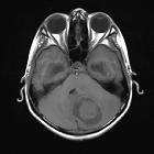
The abnormal tissue involves the cerebellar cortex and is usually confined to one hemisphere, occasionally extending to the vermis but only rarely extending to the contralateral hemisphere .
CT
- may show a non-specific hypoattenuating cerebellar mass
- calcification is sometimes seen
MRI
Widened cerebellar folia with a striated/tigroid appearance. Also described as "corduroy/laminated" appearance.
- T1: hypointense
- T2: hyperintense with apparently preserved cortical striations
- DWI: similar to normal cortex
- may show hyperintensity due to T2 shine through effect
- T1 C+ (Gd)
- enhancement is rare
- if present usually superficial, possibly due to vascular proliferation
- MR spectroscopy
- elevated lactate
- slightly reduced NAA (by about 10%)
- reduced myo-inositol (by 30-80%)
- reduced choline (by 20-50%)
- reduced Cho/Cr ratio
PET/SPECT
- FDG-PET: shows increased uptake
- Tl- SPECT: shows increased uptake
Treatment and prognosis
The dysplastic mass grows very slowly, and initial treatment revolves around treating hydrocephalus. Surgical resection is often curative, with only a few case reports of recurrence . Importantly it is crucial to remember the association with Cowden syndrome, hence, increased risk of other neoplasms such breast, endometrial and thyroid cancers. Therefore, a recommendation for further imaging or clinical assessment of possible tumors in these locations should be included in the radiologist's report.
History and etymology
It is named after Jacques Jean Lhermitte (1877-1959), a French neurologist and neuropsychiatrist, and P Duclos, who first described the condition in 1920 .
Differential diagnosis
The appearance is very characteristic and usually little differential exists, particularly when appearances are typical.
In the setting of sepsis or acute deterioration, one should consider cerebellitis or subacute cerebellar infarction.
The appearance may be mimicked by extensively nodular medulloblastoma (SHH molecular subgroup).
Siehe auch:
- Kleinhirntumoren
- Gangliozytom
- PTEN-Hamartom-Tumor-Syndrom
- Cerebellitis
- Lhermitte-Duclos-Cowden syndrome
- Kleinhirnasymmetrie
und weiter:


 Assoziationen und Differentialdiagnosen zu Lhermitte-Duclos-Syndrom: