Cisterna magna

Cisterna
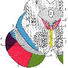
magna • Cisterns and CSF flow - Gray's anatomy illustration - Ganzer Fall bei Radiopaedia

Cisterna
magna • Cerebellomedullary cisterns (illustration) - Ganzer Fall bei Radiopaedia

Sagittalschnitte
durch Hirnstamm und Kleinhirn mit den subarachnoidalen Zisternen

Scheme of
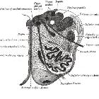
roof of fourth ventricle. The arrow is in the foramen of Magendie.

Cisterna
magna • Subarachnoid cistern (illustration) - Ganzer Fall bei Radiopaedia
The cisterna magna (also known as the cerebellomedullary cistern) is the largest of the CSF-filled subarachnoid cisterns.
Gross anatomy
The cisterna magna is located between the cerebellum and the dorsal surface of the medulla oblongata at and above the level of the foramen magnum. CSF produced in the ventricular system drains into the cisterna magna from the fourth ventricle via the median aperture (of Magendie) and the lateral apertures (of Luschka) .
Several vessels and nerves course through the lateral part of the cistern:
- vertebral arteries
- posterior inferior cerebellar arteries (PICA)
- glossopharyngeal nerve (CN IX)
- vagus nerve (CN X)
- spinal accessory nerve (CN XI)
Radiographic features
Antenatal ultrasound
- cisterna magna normally measures between 2-10 mm in the second and third trimesters
CT/MRI
- cisterna magna normally measures 3-8 mm in the midsagittal plane when measured from the posterior margin of the foramen magnum to the caudal margin of the inferior vermis
Variant anatomy
Siehe auch:
- Mega Cisterna magna
- Cerebellum
- Pons
- Medulla oblongata
- interpeduncular cistern
- vierter Ventrikel
- Foramina Luschkae
- cerebral peduncles
- pontine cistern
- subarachnoidale Zisternen
- Foramen Magendii
- Hirnventrikel
und weiter:

 Assoziationen und Differentialdiagnosen zu Cisterna magna:


