teleangiektatisches Osteosarkom


Telangiectatic osteosarcomas (TOS) are an uncommon variant of osteosarcoma that represent 2.5-12% of all osteosarcomas.
Epidemiology
Telangiectatic osteosarcomas have similar demographics to that of conventional osteosarcoma and typically presents in adolescents and young adults (reported age range of 3-67 years with a mean age of 20 years). There is a recognized male predilection.
Pathology
Most osteosarcomas have a small telangiectatic component but in order to classify as telangiectatic osteosarcoma, the telangiectatic component should comprise >90% .
Macroscopic appearance
Most of the tumor comprises of large blood-filled spaces separated by thin bony septations.
Histology
Microscopically the tumor consists of vascular sinusoids surrounded by thin septa, osteoid matrix and cells with significant pleomorphism and high mitotic rate.
Associations
Telangiectatic osteosarcoma can be a secondary lesion (arising in association with fibrous dysplasia, Paget disease, or post-radiation therapy).
Location
Most common locations by site are:
- around knee:
- femur: 50-62%
- tibia: 10-25%
- humerus: 12-16%
In terms of location within bone:
- metaphyseal: ~80%
- diaphyseal: ~20%
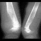
Radiographic features
Plain radiograph
Typically seen as an expansile lytic bone lesion at the metaphysis:
- geographic bony destruction with wide zone of transition tends to be more common than permeative bony destruction
- less osteoid matrix than conventional type
- pathological fractures are frequent
CT
- low attenuating fluid-fluid levels within the lesion (in ~80% of cases)
- useful at assessing associated cortical destruction
- thick peripheral and nodular septal enhancement
- osteoid matrix mineralization
MRI
- commonly shows fluid-fluid levels within the lesion (~90% of cases) with variable signal intensity
- allows appreciation of surrounding soft tissue components
- signal characteristics are often heterogeneous
- enhancement of septa with nodularity as well as the soft tissue component may be observed.
- hemorrhage appears as hyperintense on T1 and variable signal intensity on T2
Nuclear medicine
Overall, lesions tend to show marked but heterogeneous uptake on bone scans. May demonstrate a region of central photopenia representing a doughnut sign .
Treatment and prognosis
The treatment of telangiectatic osteosarcoma is often similar to that of conventional osteosarcoma: chemotherapy followed by wide surgical resection and limb salvage or amputation.
Survival rate of telangiectatic osteosarcoma (estimated at ~70%) is similar to that of conventional osteosarcoma.
Complications
They are associated with a high rate of pathological fractures .
History and etymology
First described by Sir James Paget (1814-1899), an English surgeon, in 1854 .
Differential diagnosis
Considerations on plain film include:
- aneurysmal bone cyst (ABC): should not have any soft tissue component outside the lesion. Smooth septal and rim enhancement is seen without any nodularity and without any osteoid matrix mineralization or aggressive features like cortical destruction and infiltration into surrouding tissue (unlike telangiectatic osteosarcoma).
- giant cell tumor of bone: No soft tissue component.
- osteolytic metastatic bone lesions: should not have any fluid levels
Siehe auch:
- Morbus Paget des Knochens
- Fibröse Dysplasie
- Aneurysmatische Knochenzyste
- Osteosarkom
- Riesenzelltumor des Knochens
und weiter:


 Assoziationen und Differentialdiagnosen zu teleangiektatisches Osteosarkom:


