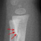
Verbiegung der langen Röhrenknochen

Röntgenbild
beider Beine bei Morbus Blount bei einem gut 1,5-jährigen Mädchen: Beidseits, links mehr als rechts, zeigt sich eine deutliche Varusdeformität mit Unregelmäßigkeit der Wachstumsfuge und Absenkung der Tibiametaphyse medial. Geringe Schrägstellung der Tibiametaphyse beidseits.


Identifier:
livinganatomypa00rotc (find matches)Title: Living anatomy and pathology;Year: 1910 (1910s)Authors: Rotch, Thomas MorganSubjects: Children Diagnosis, RadioscopicPublisher: Philadelphia London, J. B. Lippincott companyContributing Library: The Library of CongressDigitizing Sponsor: The Library of CongressView Book Page: Book ViewerAbout This Book: Catalog EntryView All Images: All Images From Book Click here to view book online to see this illustration in context in a browseable online version of this book.Text Appearing Before Image:Plate 84 PLATE 85.ADVANCED RHACHITIS. Boy, age 10 years. (Reduced 50%.) Great disturbance of the structure of the bones.Thickened cortex of concave sides of the tibiae. Pjlate 85Text Appearing After Image:PLATE 86.MARKED RHACHITIS. Boy, age 7 years. (Reduced 33£%.) Fig. 1.—Pelvis and Femora. The Roentgenograph shows especially the absorption of thelime salts in the upper epiphysis of the femur, with marked irreg-ularity in the structure of the bone, and a marked coxa vara.Note the greatly decreased density in the lower part of the fem-ora, the beak-shaped outline of the cavity of the pelvis, and thedeformed ilia with a great irregularity of the structure of the bone. A. Points to heavy deposit of cortical bone. The arrow points towards a foreign body, a needle whichwas accidentally found at the examination. Fig. 2. Photograph of the Same Subject. Shows a marked deformity from bowing of the legs. FIG. 1. Plate 86Note About Images Please note that these images are extracted from scanned page images that may have been digitally enhanced for readability - coloration and appearance of these illustrations may not perfectly resemble the original work.

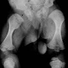
Fokale
fibrokartilaginäre Dysplasie Ulna rechts mit beginnendem Varus des Unterarm und Dezentrierung des Radiusköpfchens

Extending the
spectrum of Ellis van Creveld syndrome: a large family with a mild mutation in the EVCgene. Radiographs of patient IV-9. A) Coned epiphysis of the left second middle phalanx and prominent styloid process of the ulna B) Irregular notched tip of the distal phalanx of the hallux C) Bowing of the right humerus D) Fusion of the right proximal tibia and fibula.

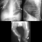
Extending the
spectrum of Ellis van Creveld syndrome: a large family with a mild mutation in the EVCgene. Radiographs of patient V-8. A) Residual appearance of postaxial polydactyly of the hand B) Abnormal appearance of some of the middle and distal phalanges of toes C) Mild lateral bowing of the humerus.
Verbiegung der langen Röhrenknochen
Siehe auch:
- Osteomyelitis
- Morbus Paget des Knochens
- Osteogenesis imperfecta
- Grünholzfraktur
- Rachitis
- Neurofibromatose Typ 1
- Achondroplasie
- Biegungsbruch
- Skelettdysplasie
- inkomplette Frakturen im Kindesalter
- Morbus Blount
- Tibiaverbiegung bei Kindern
- Skorbut
- Hypophosphatasie
- camptomelic dysplasia
- Kniest-Syndrom
- fetal limb bowing
- bone deformity from softening
- isolated congenital bowing of femur
und weiter:


 Assoziationen und Differentialdiagnosen zu Verbiegung der langen Röhrenknochen: