Osteogenesis imperfecta Typ 3

Newborn with
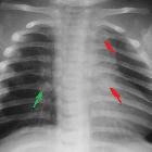
abnormally shaped extremities. Lateral radiograph of the skull (upper left) shows multiple wormian bones in the coronal and lambdoid sutures. CXR AP (upper right) shows multiple bilateral healing rib fractures. AP radiographs of the upper (lower left) and lower (lower right) extremities show multiple bilateral healing fractures of the extremities causing bowing deformities of all of the extremities.The diagnosis was osteogenesis imperfecta Type III progressive deforming.

Newborn with
abnormally shaped extremities. AP and lateral radiographs of the skull (above) show multiple wormian bones in the coronal and lambdoid sutures. CXR AP (lower left) shows multiple bilateral acute rib fractures. AP radiograph of the lower extremities (lower right) shows multiple bilateral acute and chronic fractures of the bilateral femora and tibiae and fibulae causing bowing deformities of the lower extremities.The diagnosis was osteogenesis imperfecta Type III progressive deforming.

Osteogenesis
imperfecta type II. Type III OI in a 1 year-old male. Note osteoporosis, marked deformities of the femora with bilateral fracture sequelae of the femoral diaphyses. There is hypertrophic callus at the right femoral diaphysis (arrow).

Osteogenesis
imperfecta type II. Type III OI. Note popcorn calcifications of the proximal humeri (arrows) and severe kyphoscoliosis.

Osteogenesis
imperfecta type II. Table showing the different types of OI according to the Sillence and Glorieux classification and most characteristic (imaging) features.
 Assoziationen und Differentialdiagnosen zu Osteogenesis imperfecta Typ 3:


