paranasal sinus osteoma


Osteoma of the paranasal sinuses is a common benign tumor, usually found incidentally.
Epidemiology
Osteomas are commonly found in patients undergoing imaging of the sinuses, appearing in up to 3% of CT examinations of the paranasal sinuses . They are most frequently diagnosed in 20-50 years olds, and there is a male predilection (M:F = 1.5-2.6:1) .
Clinical presentation
Most osteomas are asymptomatic and are found incidentally when imaging the sinuses either for sinonasal symptoms or for unrelated complaints.
Osteomas may become symptomatic in one of two ways:
Three possible mechanisms for pain are suggested: local effect, referred pain via the trigeminal nerve, and a prostaglandin E-2 mediated mechanism .There can be a significant inversely proportional discrepancy between the size of the lesion and the symptoms; do not simply assume because the lesion is small it does not account for the patient's symptoms.
Some osteomas are large and exophytic. They may be palpable (as is the case with skull vault osteomas) or compress structures, such as the content of the orbit . Rarely an osteoma may encroach upon the brain, and may even result in erosion of the dura with resultant CSF leak, pneumocephalus or intracranial infection (meningitis, cerebral abscess) .
More frequently they may impair normal drainage of one or more paranasal sinuses thereby resulting in acute or chronic sinusitis or even mucocele formation .
Pathology
Location
Osteomas are frequently seen elsewhere in the head and neck, particularly the mandible and outer table of the skull vault. There is a particular frequency distribution within the paranasal sinuses :
- frontal sinuses: 80%
- ethmoid air cells: ~15%
- maxillary sinuses: ~5%
- sphenoid sinus: rare
Associations
There is a well-recognized association with Gardner syndrome . Approximately 30% of patients have a history of rhinosinusitis, although a causal link has not been established .
Subtypes
Osteomas are, as the name suggests, osteogenic tumors composed of mature bone. Three histological patterns are recognized :
- also known as eburnated osteoma
- most common
- dense bone lacking Haversian system
- also known as osteoma spongiosum
- resembles 'normal' bone, including trabecular bone often with marrow
- a mixture of ivory and mature histology
Radiographic features
The radiographic appearance is that of a dense well-circumscribed mass. Ivory osteomas are uniformly very dense, whereas mature osteomas may resemble 'normal' bone with marrow space sometimes visible.
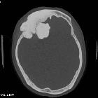
CT
CT demonstrates a well-circumscribed mass of variable density, varying from very dense (similar in density to normal cortical bone) to less dense with a ground-glass appearance. They are seen either with a sinus or less commonly exophytically growing out of a sinus.
MRI
On MRI, ivory osteomas demonstrate low signal intensity on all sequence. Mature osteomas may demonstrate some marrow signal but are also predominantly low signal on all sequences.
Treatment and prognosis
In asymptomatic cases excision is not necessarily indicated, and management varies from surgeon to surgeon. If sinonasal symptoms are present, then they can initially be managed medically (as if the osteoma is not present). In cases where the osteoma is thought to be responsible for symptoms (e.g. mucocele) then resection is required. Some surgeons prefer to excise all osteomas.
Excision may be performed either endoscopically or externally.
Differential diagnosis
General imaging differential considerations include:
- fibrous dysplasia: especially in less dense ground-glass osteomas
- other osteogenic tumors
- osteoblastoma
- osteosarcoma
- more frequently of the maxilla (rather than maxillary sinus or mandible)
- younger patients
- more aggressive appearance and rapid growth
- cemento-ossifying fibroma
- usually of the alveolar portions of the mandible or maxilla
Siehe auch:
- Osteom
- Osteosarkom
- Osteom Sinus frontalis
- kraniofaziale fibröse Dysplasie
- Osteoblastom
- cemento-ossifying fibroma
- Rhinolith
- Osteom Sinus ethmoidalis
- Osteoidfibrom der Nasennebenhöhlen
- ivory exostosis
- Osteom des Sinus maxillaris
und weiter:


 Assoziationen und Differentialdiagnosen zu Osteom NNH:


