leptomeningitis


Teenager with
left orbital and forehead swelling. Sagittal CT with contrast of the brain shows frontal and left maxillary sinusitis and extensive soft tissue swelling anterior to the forehead (upper left) and destruction of the anterior left frontal bone in the frontal sinus and extensive soft tissue swelling anterior to the left orbit (upper right). Coronal (lower left) and sagittal (lower right) T1 MRI with contrast of the brain shows diffuse meningeal enhancement, subdural empyemas along the falx and both cerebral convexities, and multiple large non-enhancing subgaleal fluid collections in the left scalp.The diagnosis was meningitis, subdural empyema, and Pott puffy tumor as complications of sinusitis.

Leptomeningitis
• Meningitis - Ganzer Fall bei Radiopaedia

Leptomeningitis
• Viral meningitis and papilledema - Ganzer Fall bei Radiopaedia

Leptomeningitis
• Bacterial meningitis - septation of subarachnoid spaces - Ganzer Fall bei Radiopaedia

Leptomeningitis
• Tubercular meningitis with tuberculomas - Ganzer Fall bei Radiopaedia

Leptomeningitis
• Meningitis from mastoiditis resulting in death - Ganzer Fall bei Radiopaedia

CNS
infectious diseases • Cryptococcal meningitis - Ganzer Fall bei Radiopaedia

Former
premature newborn, now 1 month old with seizures. Coronal US of the brain (above) shows a dilated ventricular system that contains complex echogenic fluid. Axial CT without contrast of the brain (below) shows a dilated ventricular system that contains complex fluid. The diagnosis was meningitis due to E. coli resulting in pus in the ventricular system causing communicating hydrocephalus.

Infant with
sepsis. Axial (left) and coronal (right) T1 MRI with contrast of the brain show diffuse meningeal enhancement along with a large variably enhancing solid right occipital lesion that has surrounding edema.The diagnosis was meningitis and cerebritis due to Group B streptococcus.

Ureaplasma
parvum meningitis following atypical choroid plexus papilloma resection in an adult patient: a case report and literature review. A Enhanced preoperative MRI showing a left temporal lobe-lateral ventricle tumor. B CT scans on the first day after surgery demonstrating that the tumor had been removed and replaced with CSF, with the opening of the left lateral ventricles. C Enhanced MRI images on the 43rd day after surgery demonstrating complete tumor resection
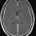
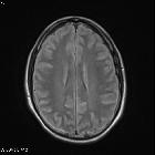
Leptomeningitis, which is more commonly referred to as meningitis, represents inflammation of the subarachnoid space (i.e. arachnoid mater and pia mater) caused by an infectious or noninfectious process.
Pathology
Etiology
Infective
- pyogenic meningitis
- elderly
- Streptococcus pneumoniae
- Listeria monocytogenes
- Neisseria meningitidis
- Gram negative bacilli
- adults
- Streptococcus pneumoniae
- Neisseria meningitidis
- Group B streptococcus
- children
- Neisseria meningitidis
- infants
- Streptococcus pneumoniae
- Neisseria meningitidis
- neonates
- Group B streptococcus
- Escherichia coli
- Listeria monocytogenes
- elderly
- viral meningitis
- Enterovirus
- JC virus meningitis
- mycobacterial meningitis
- Mycobacterium tuberculosis
- fungal meningitis
- Cryptococcus neoformans (in patients with AIDS)
- Coccidioides immitis
For a further discussion related to other etiological agents and other infective processes in the CNS, please refer to CNS infectious diseases.
Aseptic meningitis
- leptomeningeal carcinomatosis
- rheumatoid arthritis
- granulomatous
- iatrogenic
- postoperative
- hydrogel-coated aneurysm coils
Radiographic features
CT
- may be normal
- subtle hydrocephalus
- hyperdensity around basal cisterns (especially in tuberculosis)
- leptomeningeal enhancement
- complications or sources of the meningitis
MRI
- T1: may be normal; sulci may appear less hypointense than normal
- T1 C+ (Gd): leptomeningeal enhancement
- FLAIR: demonstrates hyperintense signal in CSF space, especially in the sulci
- FLAIR C+ (Gd): has shown to be more sensitive and specific than T1 C+ (Gd) sequence in spotting leptomeningeal enhancement
- MR angiography: arterial narrowing or occlusion
Treatment and prognosis
Complications
- hydrocephalus
- subdural empyema
- epidural empyema
- cerebritis and cerebral abscess
- infarction
- ventriculitis
- dural sinus thrombosis
The complications of meningitis can be remembered using the mnemonic HACTIVE.
See also
Siehe auch:
- Meningeosis neoplastica
- Sarkoidose
- Hydrocephalus
- Sinusthrombose
- Tuberkulose des ZNS
- Liquorunterdrucksyndrom
- Abszess
- leptomeningeale Kontrastmittelanreicherungen
- Ventrikulitis
- bakterielle Meningitis
- tuberkulöse Meningitis
- Pneumokokkenmeningitis
- Meningitis Ultraschall
- Cryptococcus neoformans
und weiter:
- subdurales Hygrom
- meningeale Kontrastmittelaufnahme
- verdickter Hypophysenstiel
- Normaldruckhydrozephalus
- Orbitabodenfraktur
- neuroradiologisches Curriculum
- chronisch adhäsive Arachnopathie
- subdurales Empyem
- Aquäduktstenose
- Verschlusshydrocephalus
- Mondini-Dysplasie
- Mumps
- intraspinale zystische Läsionen
- akute Otomastoiditis
- Gradenigo-Syndrom
- akute Sinusitis
- subarachnoid FLAIR hyperintensity
- pott puffy tumour
- Temporallappenepilepsie
- spinale Meningeosis neoplastica
- intrakranielle Infektionen
- communicating obstructive hydrocephalus
- kongenitale Aquäduktstenose
- Bluthirnschranke
- brudzinski sign
- multizystische Encephalopathie nach Meningitis
- meningeale Kryptokokkose
- ependymal enhancement
- hemophilus influenza B meningitis
- group B streptococcus meningitis
- Pyramidenspitzen-Osteomyelitis
- Meningoenzephalitis
- Hydrocephalus communicans
- diffusion MRI of acute cerebral infarction following meningitis

 Assoziationen und Differentialdiagnosen zu Meningitis:


