Ventrikulitis


Ventriculitis (plural: ventriculitides) refers to inflammation, usually due to infection, of the ependymal lining of the cerebral ventricles. It is most often due to meningitis.
Terminology
The entity or closely related variants have also been variously referred to as ependymitis, ventricular empyema, intraventricular abscess, and pyocephalus .
Epidemiology
Its epidemiology is varied and depends on the underlying cause.
Pathology
Etiology
- meningitis (both pyogenic and viral): most common
- cerebral abscess
- shunt/EVD related
- trauma
- CSF leakage
- a complication of intrathecal chemotherapy and neurosurgery
Meningitis is the most common underlying condition responsible for the ventriculitis. It is directly related to lower host immunity and higher virulence of the causative organism. The occurrence of direct haematogenous spread to the choroid plexus has also been suggested. In long-standing cases (>2 months) ventricular septations develop which result in compartmentalization and multiloculated hydrocephalus and makes prognosis worse.
Radiographic features
Ultrasound
Neonatal cranial ultrasound can show increased periventricular echogenicity and irregularity of the ventricular surface. Choroid plexus irregularity (thought to be due to haematogenous spread via the choroid plexus) can also be seen. Additionally, echogenic intraventricular debris may be present. When present, intraventricular septations may be well delineated on ultrasound .
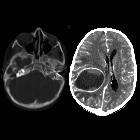
CT
Non-contrast CT of the brain usually demonstrates only non-specific features, most frequently dependent hyperdense layering material, particularly in the occipital horns of the lateral ventricles .
Hydrocephalus and periventricular low density (which probably represents reactive edema rather than transependymal edema related to hydrocephalus ) are also frequently present, as may also be the features of the underlying abnormality (e.g. meningitis, shunt/EVD, trauma)
Following administration of contrast, thin uniform enhancement of the ependymal lining of the ventricles may be seen.
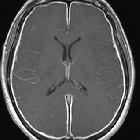
MRI
MRI demonstrates the same features as CT, with layering debris in the occipital horns frequently seen. There often an intense restricted diffusion of these intraventricular debris, as seen in the center of a brain abscess.
MRI is also more sensitive to the often subtle periventricular abnormal signal (high T2) and thin contrast enhancement.
Additionally, the periventricular region may demonstrate restricted diffusion on DWI and ADC.
Differential diagnosis
The main differential diagnosis is that of ependymal lining enhancement, which includes ependymal spreading of glioblastoma or primary CNS lymphoma. In these cases, the enhancement is, usually, bumpy/nodular. Extracranial neoplasm metastases and germinoma may also be responsible for similar findings.
Siehe auch:
- ependymitis granularis
- Hirnabszess
- Meningitis
- Seitenventrikel
- Hirnventrikel
- transependymales Ödem
- bakterieller Hirnabszess
- Periventrikulitis
und weiter:


 Assoziationen und Differentialdiagnosen zu Ventrikulitis: