ependymitis granularis

Ependymitis
granularis • Ependymitis granularis - Ganzer Fall bei Radiopaedia
Ependymitis
granularis • Ependymitis granularis - Ganzer Fall bei Radiopaedia
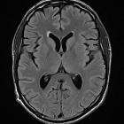
Ependymitis granularis sounds far more worrying than it actually is. The term refers to symmetrical foci of periventricular high T2 and FLAIR signal hyperintensity anterior and lateral to the frontal horns. It is just an anatomical variant, usually small, less than 1 cm, and has a triangular morphology extending laterally from the callosal genu. Pathologic lesions tend to be larger and demonstrate corresponding low signal intensity in T1WI.
Pathology
Three findings contribute to the appearance :
Despite the name, which suggests an inflammatory cause, and a 1926 article that claimed it as a cause of chronic internal hydrocephalus, it is just an anatomical variant.
Differential diagnosis
Siehe auch:


 Assoziationen und Differentialdiagnosen zu ependymitis granularis:
