accessory ossicles of the foot

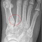
Kleines Os
intermetatarseum im Röntgenbild: Zusätzlicher Knochen zwischen den Basen des ersten und zweiten Mittelfußknochens. Zufallsbefund.


Accessory
ossicles of the foot • Os subfibulare - Ganzer Fall bei Radiopaedia


Os vesalianum
pedis • Accessory ossicles of the foot (illustration) - Ganzer Fall bei Radiopaedia

Accessory
ossicles of the foot • Os vesalianum pedis - Ganzer Fall bei Radiopaedia

Identifier:
americanjournroen09ameruoft (find matches)Title: The American journal of roentgenology, radium therapy and nuclear medicineYear: 1906 (1900s)Authors: American Radium Society American Roentgen Ray SocietySubjects: Radiotherapy X-raysPublisher: Springfield, Ill. C.C. ThomasContributing Library: Gerstein - University of TorontoDigitizing Sponsor: University of TorontoView Book Page: Book ViewerAbout This Book: Catalog EntryView All Images: All Images From Book Click here to view book online to see this illustration in context in a browseable online version of this book.Text Appearing Before Image:ut yet so clearlj marked off on thesurface that there is no question as to itsidentity. Only when it is so completelyfused with another bone that it loses itsown characteristics is its nature in doubt. In order to gain a better idea as to thenature of these bones, I have madecomposite drawings oi the toot of anadult, both dorsal and plantar views,which show the supernumerary ossicles intheir usual relationships to the constantelements. The variations in size, shape,and position of the individual ossiclesare not of sufficient degree to cause anydifliculu in their recognition when seen the Si) rgi G< 1 ral. The Supernumerary Pedal Bones 45 in the macerated foot or in the roentgeno-gram, once their general characteristics ind (Figs. 1 in m and positions are fixedand 2). The following is a tabulation of all thesupernumerary ossicles which, up to the 4. Cuboideum (a) cuboideum secondarium 5. Cuneiforme primum (a) cuneiforme I bipartitum 6. Cuneiforme secundum (a) intercuneiformeText Appearing After Image:Fig. 1. Dorsal view. Composite foot showing all knownsupernumerary bones. present time, have been found on dis-section and proved to be true bony vari-ants. To this there is but one exception,and that is the secondary astragaluswhich was seen in a stereoscopic picture,and which the writer has described inanother publication. With this one excep-tion all the bones are firmly established astrue supernumerary ossicles. 1. Astragalus (a) trigonum (6) astragalus secondarius 2. Calcaneus (a) os sustentaculi (b) calcaneum secondarium 3. Naviculare (a) tibiale externumi/m gupranaviculare Fig. 2. Plantar view. (Same foot as Fig. I.) Cuneiforme tertium (a) os unci (processus uncinatus cunei- form is) (b) Intermetatarseum (c) Os vesalianum (d) Os peroneum Normal bonesSupernumerary ossicles Total <3 20 Trigonum. This is the best known of thesupernumerary tarsal bones, and is probablythe one most intensively studied. The firstaccurate description was by Schwegel in1858, although it was first reNote About Images Please note that these images are extracted from scanned page images that may have been digitally enhanced for readability - coloration and appearance of these illustrations may not perfectly resemble the original work.


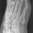
Kleines Os
intermetatarseum zwischen den Basen von Metatarsale eins und zwei als Zufallsbefund im Röntgenbild des Fußes.

X-ray of the
left ankle by anteroposterior projection, with areas representing where common accessory bones would be, colored by their prevalences, and with representative shapes.


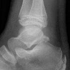
Knochenkern
dorsal am Talus bei einem 10-Jährigen in der Position eines Os trigonums.


Location of
the accessory ossicles of the tarsus. On the left, the plantar aspect of the right foot, on the right the dorsal aspect of the right foot. The more dorsally situated ossicles are shown in closed circles on the right and open circles on the left. The ossicles lying closer to the plantar surface of the foot (the sole) are shown in closed circles on the left and open circles on the right. The half-open circles are situated even more dorsally then the others, being situated just below the medial and lateral malleolus. 1: Os cuneometatarsale I plantare, 2: os uncinatum, 3: os sesamoideum tibialis posterior, 4: os sesamoideum peroneum, 5: os cuboideum secundarium, 6: os trochleare calcanei, 7: os in sinus tarsi, 8: os sustentaculum tali, 9: os talocalcaneale posterius, 10: os aponeurosis plantaris, 11: os subcalcaneum, 12: os sesamoideum tibialis anterior, 13: os cuneometatarsale I tibiale, 14: os intermetatarsale I, 15: os cuneometatarsale II dorsale, 16: os paracuneiforme, 17: os cuneonaviculare, 18: os intercuneiforme, 19: os intermetatarsale IV, 20: os talonaviculare, 21: os vesalianum pedis, 22: os tibiale externum, 23: os talotibiale dorsale, 24: os supratalare, 25: os calcaneus secundarius, 26: os subtibiale, 27: os subfibulare, 28: os retinaculi, 29: os calcaneus accessorius, 30: os trigonum, 31: os supracalcaneum, 32: os tendinis calcanei

Bipartite
medial cuneiform: congenital variant in a patient with foot trauma. MRI findings. Fig. 3: sagittal T1 showing dorsal and plantar segments forming the "E" sign.

Accessory
ossicles of the foot • Accessory ossicles of the foot (illustration) - Ganzer Fall bei Radiopaedia

Accessory
ossicles of the foot • Accessory ossicles of the foot - Ganzer Fall bei Radiopaedia

Accessory
ossicles of the foot • Os tibiale externum - Ganzer Fall bei Radiopaedia

Accessory
ossicles of the foot • Os peroneum - Ganzer Fall bei Radiopaedia

Accessory
ossicles of the foot • Os trigonum - Ganzer Fall bei Radiopaedia

Accessory
ossicles of the foot • Os supranaviculare - Ganzer Fall bei Radiopaedia

Accessory
ossicles of the foot • Os infranaviculare - Ganzer Fall bei Radiopaedia

Sesamoid •
Articulating os intermetatarseum - Ganzer Fall bei Radiopaedia
Accessory ossicles of the feet are common developmental variants with almost 40 having been described. Some of the more common include :
- os peroneum
- os subfibulare
- os subtibiale
- os tibiale externum (accessory navicular)
- os trigonum
- os calcaneus secundaris
- os intermetatarseum
- pars peronea metatarsalis primi (pars peronea metatarsalia)
- os supratalare
- bipartite hallux sesamoid
- os supranaviculare
- os infranaviculare (cuneonavicular ossicle)
Knowledge of their presence is helpful so that they are not misdiagnosed as fractures.
Siehe auch:
- Akzessorische Handknochen
- Os tibiale externum
- Os peroneum
- Os trigonum
- Os subfibulare
- Os calcaneus secundarius
- Os subtibiale
- Akzessorischer Knochen
- Os intermetatarseum
- Os talotibiale
- Os cuneiforme mediale bipartitium
- Fußwurzelknochen
- akzessorisches Os cuneiforme mediale
- Pars peronea metatarsalis primi
- accessory ossification centers
und weiter:

 Assoziationen und Differentialdiagnosen zu akzessorische Knochen Fuß:


