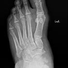
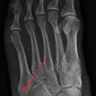
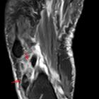
Os peroneum


Os peroneum
• Os peroneum fracture - Ganzer Fall bei Radiopaedia

Os peroneum
• Bilateral os peroneum - Ganzer Fall bei Radiopaedia

Identifier:
americanjournroen09ameruoft (find matches)Title: The American journal of roentgenology, radium therapy and nuclear medicineYear: 1906 (1900s)Authors: American Radium Society American Roentgen Ray SocietySubjects: Radiotherapy X-raysPublisher: Springfield, Ill. C.C. ThomasContributing Library: Gerstein - University of TorontoDigitizing Sponsor: University of TorontoView Book Page: Book ViewerAbout This Book: Catalog EntryView All Images: All Images From Book Click here to view book online to see this illustration in context in a browseable online version of this book.Text Appearing Before Image:ut yet so clearlj marked off on thesurface that there is no question as to itsidentity. Only when it is so completelyfused with another bone that it loses itsown characteristics is its nature in doubt. In order to gain a better idea as to thenature of these bones, I have madecomposite drawings oi the toot of anadult, both dorsal and plantar views,which show the supernumerary ossicles intheir usual relationships to the constantelements. The variations in size, shape,and position of the individual ossiclesare not of sufficient degree to cause anydifliculu in their recognition when seen the Si) rgi G< 1 ral. The Supernumerary Pedal Bones 45 in the macerated foot or in the roentgeno-gram, once their general characteristics ind (Figs. 1 in m and positions are fixedand 2). The following is a tabulation of all thesupernumerary ossicles which, up to the 4. Cuboideum (a) cuboideum secondarium 5. Cuneiforme primum (a) cuneiforme I bipartitum 6. Cuneiforme secundum (a) intercuneiformeText Appearing After Image:Fig. 1. Dorsal view. Composite foot showing all knownsupernumerary bones. present time, have been found on dis-section and proved to be true bony vari-ants. To this there is but one exception,and that is the secondary astragaluswhich was seen in a stereoscopic picture,and which the writer has described inanother publication. With this one excep-tion all the bones are firmly established astrue supernumerary ossicles. 1. Astragalus (a) trigonum (6) astragalus secondarius 2. Calcaneus (a) os sustentaculi (b) calcaneum secondarium 3. Naviculare (a) tibiale externumi/m gupranaviculare Fig. 2. Plantar view. (Same foot as Fig. I.) Cuneiforme tertium (a) os unci (processus uncinatus cunei- form is) (b) Intermetatarseum (c) Os vesalianum (d) Os peroneum Normal bonesSupernumerary ossicles Total <3 20 Trigonum. This is the best known of thesupernumerary tarsal bones, and is probablythe one most intensively studied. The firstaccurate description was by Schwegel in1858, although it was first reNote About Images Please note that these images are extracted from scanned page images that may have been digitally enhanced for readability - coloration and appearance of these illustrations may not perfectly resemble the original work.


Os peroneum
• Os peroneum - diagram - Ganzer Fall bei Radiopaedia

Accessory
ossicles of the foot • Os peroneum - Ganzer Fall bei Radiopaedia

Os peroneum
• Os peroneum with os tibiale externum - Ganzer Fall bei Radiopaedia

Mehrere
Beispiele für ein Os peroneum im schrägen Röntgenbild des Fußes mit typischer Lage und Größe. (Beachte auch die Gicht im linken Teilbild.)

Os peroneum
• Os peroneum - Ganzer Fall bei Radiopaedia

Os peroneum
• Peroneus longus tendon rupture associated with an os peroneum - Ganzer Fall bei Radiopaedia

Os peroneum
• Os peroneum - Ganzer Fall bei Radiopaedia

Os peroneum
• Os peroneum - Ganzer Fall bei Radiopaedia

Os peroneum
• Os peroneum - Ganzer Fall bei Radiopaedia

Os peroneum
• Freiberg disease - 2nd metatarsal - Ganzer Fall bei Radiopaedia

Avulsion
fracture of the 5th metatarsal styloid • Pseudo-Jones fracture - Ganzer Fall bei Radiopaedia

Avulsion
injury • Osteomyelitis and avulsion fracture in a diabetic foot - Ganzer Fall bei Radiopaedia

Os peroneum
• Os intermetatarseum - Ganzer Fall bei Radiopaedia
An os peroneum is a small accessory bone located at the lateral plantar aspect of the cuboid within the substance of the peroneus longus tendon as it arches around the cuboid. It is a very common anatomical variant, seen in up to 26% of feet .
Differential diagnosis
It should not be mistaken for:
- os vesalianum
- os cuboideum secundarium
- apophysis of the 5 metatarsal
- avulsion fracture (pseudo-Jones fracture)
Related pathology
It can produce pathology (os peroneum syndrome) including fracture, peroneus longus tenosynovitis and peroneus longus tendon tear.
Siehe auch:
- akzessorische Knochen Fuß
- Avulsionsfraktur Basis Metatarsale 5
- Ruptur der Sehne des Musculus peroneus longus
- Musculus peroneus longus
- geteiltes Os peroneum
- Apophysitis an der Basis des Os metatarsale 5 (Iselin-Krankheit)
- Os peroneum nach proximal verlagert
- Fraktur Os peroneum
- apophysis of the 5th metatarsal
und weiter:

 Assoziationen und Differentialdiagnosen zu Os peroneum:


