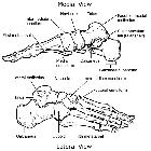
Fraktur Os cuboideum

Update on
diagnosis and management of cuboid fractures: Sagittal view of the right ankle obtained via magnetic resonance imaging. Undisplaced cuboid fracture extending to the middle of the calcaneocuboid joint.

A case of
cuboid bone stress fracture in a senior high school rugby athlete: Fig. 2. Magnetic resonance imaging findings. (A) Axial T1-weighted image shows continuous hypointense signals (arrow) from the cuneiform joint surface of the cuboid bone toward the lateral side. (B) Fat suppression T2-weighted image shows hyperintense signals (arrow).
Fraktur Os cuboideum
Siehe auch:
und weiter:


 Assoziationen und Differentialdiagnosen zu Fraktur Os cuboideum: