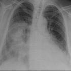
air bronchogram


Air bronchogram refers to the phenomenon of air-filled bronchi (dark) being made visible by the opacification of surrounding alveoli (grey/white). It is almost always caused by a pathologic airspace/alveolar process, in which something other than air fills the alveoli. Air bronchograms will not be visible if the bronchi themselves are opacified (e.g. by fluid) and thus indicate patent proximal airways.
Air bronchograms can be seen with several processes:
- pulmonary consolidation
- pulmonary edema: especially with alveolar edema
- non-obstructive atelectasis
- severe interstitial lung disease
- neoplasms: bronchioloalveolar carcinoma; pulmonary lymphoma
- pulmonary infarct
- pulmonary hemorrhage
- normal expiration
Air bronchograms that persist for weeks despite appropriate antimicrobial therapy should raise the suspicion of a neoplastic process. CT may be planned in such cases.
Radiographic features
Ultrasound
Sonographic air bronchograms arise as a secondary consequence of an extreme perturbation of the air-fluid relationship in the lung parenchyma, in which fluid-filled alveoli act as an excellent acoustic medium and allow visualization of the lung parenchyma. Arborising tubular structures representing the bronchial tree may be visualized which, when patent, appear to contain punctiform-to-linear foci. These structures may remain fixed in position throughout the respiratory cycle or be observed to propagate distally and proximally with inspiration and expiration, respectively. This distinction is important for determining the etiology of the underlying pathology ;
- dynamic air bronchograms move centrifugally with respiration
- represent fluid mixed with air inside larger bronchi, which are in continuity with the gas inspired by the patient
- indicates a non-retractile consolidation, ruling out resorption atelectasis
- the specificity of 94% and a positive predictive value of 97% for pneumonia as the cause of the consolidation
- static air bronchograms lack detectable movement
- indicate isolated, trapped air, consistent with resorptive atelectasis
See also
Siehe auch:
- Atelektase
- Konsolidierung der Lunge
- Lungenödem
- Pneumonie
- In Situ Adenokarzinom der Lunge
- Interstitielle Lungenerkrankung
- Lymphom pulmonale Manifestation
- Lungeninfarkt
- airspace disease
und weiter:


 Assoziationen und Differentialdiagnosen zu Bronchopneumogramm: