Angiomatose der Mamma

Angiomatosis
of breast with osseous metaplasia. Screening mammography of the left breast (Craniocaudal view) showing focal asymmetry with specks of punctate calcification in the outer quadrant of the left breast.
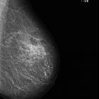
Angiomatosis
of breast with osseous metaplasia. Screening mammography of the left breast (mediolateral oblique view) showing focal asymmetry with specks of punctate calcification in the upper quadrant of the left breast.

Angiomatosis
of breast with osseous metaplasia. Screening mammography of the left breast (spot compression view) showing focal asymmetry with specks of punctate calcification (arrow) in the left breast.

Angiomatosis
of breast with osseous metaplasia. Ultrasonography of the left breast showing a cluster of low echogenic area intermingled with normal parenchyma in the retroglandular region in the upper outer quadrant of the left breast.

Angiomatosis
of breast with osseous metaplasia. Colour Flow Doppler of the lesion in the upper outer quadrant of the left breast showing vascularity in the tubular low echogenic area.

Angiomatosis
of breast with osseous metaplasia. Magnetic Resonance T1 weighted image showing intermediate signal intensity of the lesion in the left breast.

Angiomatosis
of breast with osseous metaplasia. Magnetic Resonance T2 weighted image showing high signal intensity of the lesion in the left breast.

Angiomatosis
of breast with osseous metaplasia. Post-contrast T1 weighted fat-suppressed image showing avid early homogeneous enhancement of the lesion.

Angiomatosis
of breast with osseous metaplasia. Post-contrast T1 weighted fat-suppressed image of the left breast showing persistent homogeneous enhancement of the lesion at 2 minutes.

Angiomatosis
of breast with osseous metaplasia. Post-contrast T1 weighted fat-suppressed image of the left breast showing persistent homogeneous enhancement of the lesion at 4 minutes.
Angiomatose der Mamma
Siehe auch:


 Assoziationen und Differentialdiagnosen zu Angiomatose der Mamma:
