Angiosarkom der Mamma


Breast angiosarcomas are a rare vascular breast malignancy.
Epidemiology
As primary tumors of the breast, they account for ~0.04% of all breast cancers and tend to occur in younger women, in their 3 to 4 decades.
Secondary angiosarcoma, related to prior therapy of breast cancer, has an estimated incidence of ~0.09-0.16% and occurs in older women (peak age 6 decade ).
Clinical presentation
The classic presentation is a painless localized blue or purple color change of the breast skin, with singular or multifocal lesions which may resemble other benign vascular lesions such as angioma or telangiectasis .
Patients may also present with swelling, a sensation of fullness, or rapid breast growth .
Pathology
The tumors are of endovascular origin. There are two main types:
- primary angiosarcoma of the breast
- secondary angiosarcoma of the breast
- radiation-induced angiosarcoma: can occur following breast irradiation
- lymphedema-associated cutaneous angiosarcoma
Location
Specific location of the tumor varies according to whether it is a primary or secondary lesion:
- primary breast angiosarcoma are thought to arise within the breast parenchyma, with secondary involvement of the skin
- secondary breast angiosarcoma mainly involve the skin, with or without involvement of underlying breast parenchyma
Associations
- prior breast irradiation: radiation-induced breast angiosarcomas tend to occur after a significant latent period post-radiation (~5 years)
- Stewart-Treves syndrome
Radiographic features
Mammography
Lesions can sometimes be occult on mammography .
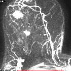
Breast MRI
A heterogeneous mass is usually seen on MRI. Reported signal characteristics include :
- T1: usually low signal intensity
- T2: usually high signal intensity
- C+ (Gd): low-grade lesions show progressive enhancement whereas high-grade lesions show rapid enhancement and washout
Treatment and prognosis
Angiosarcoma of the breast is an extremely aggressive tumor and is associated with a poor prognosis.
The tumor may be mis-diagnosed on the basis of fine-needle biopsy; thus, core biopsy is mandatory for diagnosis .
Surgical excision with wide margin is the standard of care, typically a mastectomy. Axillary nodal dissection is usually not performed, as the malignancy features haematogenous spread.
Despite aggressive surgery, there is a high local and distant recurrence rate.
Siehe auch:
und weiter:


 Assoziationen und Differentialdiagnosen zu Angiosarkom der Mamma:

