anterior interosseous nerve syndrome
Anterior interosseous nerve syndrome (AINS), also known as Kiloh-Nevin syndrome, is one of three common median nerve entrapment syndromes; the other two being pronator teres syndrome and the far more common carpal tunnel syndrome.
Epidemiology
AINS is a rare entrapment syndrome, with comparatively little robust epidemiological data. AINS is said to account for less than 1% of neuropathies of the upper limb .
Clinical presentation
AINS is a pure motor neuropathy, as the anterior interosseous nerve contains no sensory fibers; dull forearm pain is however sometimes mentioned by patients.
Typically, patients fail to make an “O.K.”-sign, as flexion of the interphalangeal joint of the thumb and the distal interphalangeal joint of the index finger is impaired.
Another sensitive test is the pinch test: a patient with AINS will also not be able to pinch a sheet of paper between his thumb and index finger, instead clamping the sheet between his extended thumb and index fingers, akin to a tong rather than a clamp. Weakness of the pronator quadratus muscle manifests itself in pronation weakness with a flexed elbow .
AINS can be confounded by the Martin-Gruber anastomosis, present in up to 25% of the population: in these cases, the anterior interosseous nerve gives off branches to the ulnar nerve, creating atypical motor innervation patterns of the forearm and hand and thus effacing the typical clinical symptoms.
Pathology
The etiology is highly debated. Two common causes of AINS are compression neuropathy and brachial plexus neuritis .
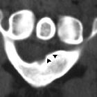
AINS can be caused by compression between the heads of the pronator teres muscle and the proximal edge of the FDS arch. The anterior interosseous nerve is furthermore prone to entrapment by anatomical variants, Gantzer’s muscle, an anoumalous head of the FPL (flexor pollicis longus muscle) , being the most noteworthy, as it is found in up to 52% of the population .
Distribution
In complete AINS, the flexor pollicis longus (FPL), the radial part (2nd and 3rd digit) of the flexor digitorum profundus (FDP) and pronator quadratus (PQ) are affected.
Radiographic features
Ultrasound and MRI are the two imaging modalities which best lend themselves to investigating entrapment syndromes. Next to directly visualizing direct causes [e.g. primary nerve or sheath tumors, ganglion cysts, osseous spurs, anatomical variants (e.g. Gantzer muscle), recognizing pathological muscle signal patterns on MRI can inversely point to the affected nerve.
MRI
Look for the pattern of muscle signal changes on fluid-sensitive (STIR, PD or T2w fat sat) sequences to uncover the affected nerve.
In AINS, the flexor pollicis longus (FPL), the radial part (2nd and 3rd digit) of the flexor digitorum profundus (FDP) and pronator quadratus (PQ) will be accordingly affected. In chronic cases, additional hyperintense signal changes can occur on T1w imaging secondary to lipomatous atrophy. Care must be taken in assessing the PQ muscle, however: hyperintense signal here has been shown to be a frequent normal finding of unclear etiology .
Although not strictly necessary in secondary entrapment syndromes (i.e. those not caused by space-occupying lesions etc.), intravenous application of contrast medium will show enhancement of denervated muscles.
Treatment and prognosis
AINS can be treated conservatively with extremity rest and NSAR; corticosteroids have been used, as well. Surgical success rates is about 73% in one series .
Differential diagnosis
Anterior interosseous nerve syndrome must be differentially diagnosed from:
- other median nerve entrapment syndromes
History and etymology
Anterior interosseous nerve syndrome (Kiloh-Nevin syndrome) was first described in 1948 by Parsonage and Turner and further defined in 1952 by Kiloh and Nevin .
See also
- median nerve entrapment syndromes
- pronator teres syndrome
- carpal tunnel syndrome
- anterior interosseous nerve syndrome
Siehe auch:


 Assoziationen und Differentialdiagnosen zu Kiloh-Nevin-Syndrom: