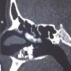
antrolith
An antrolith is a calcified mass within the maxillary sinus. The origin of the nidus of calcification may be extrinsic (foreign body in sinus) or intrinsic (stagnant mucus, fungal ball).
Most antroliths are small and asymptomatic. Larger ones may present as a chronic maxillary sinusitis with symptoms like pain and discharge.
Radiographic features
They appear as solitary or multiple, faintly to extremely radiopaque masses embedded within the mucoperiosteum. The sinus wall is intact.
Treatment and prognosis
Small antroliths require no treatment. However, larger or symptomatic masses need to be are surgically removed.
Siehe auch:
und weiter:
 Assoziationen und Differentialdiagnosen zu Rhinolith:


