Auftreibung der Rippen

The chest
radiograph in this child shows nodular prominence of the costochondral junctions, consistent with rickets.


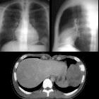
Teenager with
fever and left pleuritic chest pain. CXR PA and lateral (above) show a round mass on the lateral and middle aspect of the dome of the left hemidiaphragm. Axial CT with contrast of the chest (below) shows the soft tissue mass to be arising from a rib which has extensive and aggressive periosteal reaction.The diagnosis was Ewing sarcoma of the rib (Askin tumor).

Chondromyxoid
fibroma of rib with a novel chromosomal translocation: a report of four additional cases at unusual sites. Computed tomography scan of chest (case no. 2) demonstrating a radiolucent lesion of the right anterior second rib with cortical bubbling or blebbing, and without radiographic evidence of an acute fracture, calcification or malignancy (arrow). R indicates right side.

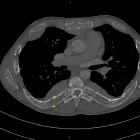
Osteoblastoma
of the rib with CT and MR imaging: a case report and literature review. CT reveals a lytic lesion in right fifth posterior rib with ossified matrix.

Tumor and
tumorlike conditions of the pleura and juxtapleural region: review of imaging findings. Diagnosis: Ewing sarcoma. Technique: standard chest radiography and contrast-enhanced chest CT. Description: A 23-year-old man was referred for a chest radiograph due to pain at the right hemithorax. The postero-anterior chest radiographs (a, b) show an opacity (arrow) projecting at the level of the 7th right rib. An external radio-opaque marker was placed on the painful area. The additional contrast-enhanced CT further characterizes the mass encasing the 7th right rib with a locoregional aggressive periosteal sunburst type reaction and a surrounding soft tissue component (c, d). On the axial T1-weighted MR image (e) the lesion has an isointense signal to muscle and a high signal intensity on the axial FS T2-WI (f) with invasion of the muscles of the chest wall (arrows). After intravenous contrast administration, the lesions show a heterogeneous enhancement (g), predominantly at the periphery of the lesion. The coronal T2-weighted MR image (h) shows the extrinsic impression of the lesion on the liver

Tumor and
tumorlike conditions of the pleura and juxtapleural region: review of imaging findings. Diagnosis: chondrosarcoma. Technique: contrast-enhanced chest CT and MRI. Description: A 64-year-old woman with a history of smoking, presented due to shortness of breath. The axial contrast-enhanced CT scan (a, b) reveals an expansile lesion with extensive calcifications located at the costochondral junction of the right 3rd rib (note a typical arc-and-ring pattern of calcification). On the axial T1-weighted MR image (c) the lesions have a high signal intensity. The axial T2-weighted MR image (d) shows a peripheral hyperintense mass with internal low-intensity foci, reflecting the cartilaginous matrix with chondroid calcifications

Fibrous
dysplasia • Fibrous dysplasia - ribs - Ganzer Fall bei Radiopaedia

Fibrous
dysplasia for radiologists: beyond ground glass bone matrix. Multimodality imaging in polyostotic fibrous dysplasia (FD). a, b PET/CT with 18-F-NaF demonstrates multiple lucent FD lesions seen on CT with corresponding areas of mild radiotracer uptake on PET. c–f Lesions demonstrate intermediate T1 signal intensity on MRI (c), intermediate-to-low signal intensity on T2 (d), slightly hyperintense signal intensity on DWI (e), uniform enhancement after contrast administration (f)

Fibrous
dysplasia for radiologists: beyond ground glass bone matrix. The significance of MRI with diffusion-weighted imaging (DWI) in the patient with polyostotic fibrous dysplasia (FD). CT (a), T2-weighted MRI (b) and DWI (c) show multiple rib lesions (green arrows). The left rib lesion (red arrow) demonstrates restricted diffusion, which requires further evaluation to rule out malignant transformation

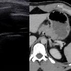
Zoledronic
acid in metastatic chondrosarcoma and advanced sacrum chordoma: two case reports. a Thoracic CT scan in the patient with chondrosarcoma shows at right the lesion involving muscles and ribs. Lung metastases were visualized. b Coronal section displays the large tumor.

Thalassemia
• Extramedullary haemopoiesis in a thalassemia major patient - Ganzer Fall bei Radiopaedia

Teenager with
respiratory distress. CXR AP shows diffuse osteopenia of the bones, lobulated masses projecting over the posterior portions of the ribs bilaterally, and an expanded appearance of the anterior and posterior aspects of the ribs.The diagnosis was thalassemia with extramedullary hematopoesis.


Intrathoracic
extramedullary haematopoiesis and skeletal involvement in a case of thalassaemia intermedia. Chest axial image shows diffuse structural remodelling of bones with destruction of trabeculae and multiple cortical interruptions; ribs appear widened (arrow).

Intrathoracic
extramedullary haematopoiesis and skeletal involvement in a case of thalassaemia intermedia. Chest axial image shows bilateral paravertebral and right pericostal well-marginated masses, demonstrating mild enhancement after intravenous injection of contrast medium; diffuse decreased density of bones with structural remodelling is also seen.

Thalassemia
• Thalassemia - Ganzer Fall bei Radiopaedia

Rachitic
rosary • Rickets - Ganzer Fall bei Radiopaedia

Toddler with
poor weight and height gain. CXR AP (above) shows prominence of the anterior ribs near the costochondral junctions (rachitic rosary). AP and lateral radiograph of the wrist (below) shows cupping of the distal radius and ulna metaphyses.The diagnosis was Vitamin D deficiency rickets.

Rickets •
Rickets - Ganzer Fall bei Radiopaedia


Jeune-Syndrom
Thorax: Verkürzung der Rippen mit schmalem Brustkorb. Verdichtungen der Lunge wohl durch Minderbelüftung.

Fibrous
dysplasia • Fibrous dysplasia of the rib - Ganzer Fall bei Radiopaedia
 Assoziationen und Differentialdiagnosen zu Auftreibung der Rippen:


