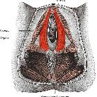
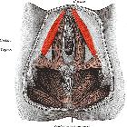
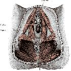
Beckenbodenmuskulatur

Dynamic
magnetic resonance imaging of the female pelvic floor—a pictorial review. Pelvic muscles. (a, b) Coronal (a) and axial (b) T2-TSE images of the pelvic diaphragm, showing the iliococcygeus (white arrows) and puborectalis (red arrows) muscles

Dynamic
magnetic resonance imaging of the female pelvic floor—a pictorial review. Pubococcygeal line (PCL). Sagittal midline T2-TSE image with the PCL represented (red line) connecting the inferior border of the pubic symphysis and the last coccygeal joint

Dynamic
magnetic resonance imaging of the female pelvic floor—a pictorial review. Anorectal junction (ARJ). (a, b) Axial T2-TSE image (a) at the plane of the puborectalis muscle (red arrows), the same plane of the ARJ. Sagittal T2-TSE image (b) showing the posterior wall of the ARJ (white arrow)

Dynamic
magnetic resonance imaging of the female pelvic floor—a pictorial review. Puborectalis defect. Axial T2-TSE image of the puborectalis muscle, demonstrating a right posterior defect, asymmetry, and atrophy, with lipomatous involution (red arrow)
Beckenbodenmuskulatur
Siehe auch:
- Musculus bulbospongiosus
- Musculus ischiocavernosus
- Musculus transversus perinei profundus
- Musculus transversus perinei superficialis
- Musculus coccygeus
- Beckenmuskulatur
- Levator ani muscles
- Musculus levator ani
- Beckenbodeninsuffizienz
- Beckenbodendysfunktion
- Musculus pubococcygeus
- Musculus puborectalis
- Musculus iliococcygeus
und weiter:

 Assoziationen und Differentialdiagnosen zu Beckenbodenmuskulatur:


