bilaterale parietale Ausdünnung Schädelkalotte

Ausdünnung
der Kalotte beidseits parietal, weitgehend symmetrisch mit Verlust der Diploe hier. Zufallsbefund bei einer 83-jährigen. (Osteoporose?) Nicht zu verwechseln mit Foramina parietalia. CT als Volumen rendering und sagittal.

Radiological
review of skull lesions. Parietal thinning. Axial (a) and coronal (b) head CT images shows thinning of the parietal bones (thick arrows)

Ausdünnung
der Kalotte beidseits parietal, weitgehend symmetrisch mit Verlust der Diploe hier. Zufallsbefund bei einer 83-jährigen. (Osteoporose?) Nicht zu verwechseln mit Foramina parietalia. CT und MRT T1w ohne KM coronar.

Ausdünnung
der Kalotte beidseits parietal, weitgehend symmetrisch mit Verlust der Diploe hier. Zufallsbefund bei einer 83-jährigen. (Osteoporose?) Nicht zu verwechseln mit Foramina parietalia. CT coronar und axial.

Parietale
Ausdünnung der Schädelkalotte wie sie meist beidseitig vorkommt, hier asymmetrisch mit links nur minimalem Befund. Computertomographie coronar, axial (sek. rekonstruiert mit Bewegungsartefakt) und Volumen Rendering (VR Ansicht von oben, so dass rechte Bildseite auch rechte Patientenseite ist).

Fokale
parietale Kalottenausdünnung unklarer Genese auf Höhe der Foramina parietalia (hier nur rechts). Computertomographie Volumen rendering (Blick von rechts dorsal) und coronare Rekonstruktion.

Imaging of
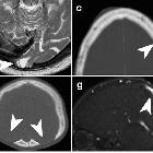
skull vault tumors in adults. Calvarial pseudolesions. NECT (a, c–f), T2WI (b), MR venography (g, h). Parasagittal occipital arachnoid granulation (a). Occipital encephalocele (b). Dilated vascular canals and lacunae (c). Hyperostosis frontalis interna (d). Bilateral thinning of the parietal bones (e). Enlarged parietal foramina (f). Congenital sinus pericranii (g). Iatrogenic acquired sinus pericranii from a prior ventricular drainage (h)

Bilateral
thinning of the parietal bones • Bilateral thinning of the parietal bones - Ganzer Fall bei Radiopaedia

Bilateral
thinning of the parietal bones • Bilateral thinning of the parietal bones - Ganzer Fall bei Radiopaedia

Bilateral
thinning of the parietal bones • Parietal skull vault thinning - Ganzer Fall bei Radiopaedia

Bilateral
thinning of the parietal bones • Biparietal osteodystrophy - Ganzer Fall bei Radiopaedia

Bilateral
thinning of the parietal bones • Biparietal osteodystrophy (thinning of parietal bones) - Ganzer Fall bei Radiopaedia

Bilateral
thinning of the parietal bones • Bilateral thinning of the parietal bones - Ganzer Fall bei Radiopaedia

Bilateral
thinning of the parietal bones • Bilateral thinning of the parietal bones - Ganzer Fall bei Radiopaedia

Biparietale
Ausdünnung der Schädelkalotte, als möglicher Hinweis auf Osteoporose.
 nicht verwechseln mit: Foramina parietalia permagna
Bilateral thinning of the parietal bones, also known as biparietal osteodystrophy, is an uncommon, slowly progressive acquired disease of middle-aged people with slight female predilection. It is typically an incidental finding.
Pathology
The etiology is unknown but is thought to be an age-related process due to osteoporosis.
Microscopic appearance
On histologic examination, there is a lack of osteoclasts.
Radiographic features
Nuclear medicine
Bone scan usually shows well defined areas of photopenia, which further supports the osteoporotic etiology of this process.
Treatment and prognosis
In severe cases where brain is exposed to atmospheric pressure, cranioplasty may be indicated.
Siehe auch:
und weiter:

 Assoziationen und Differentialdiagnosen zu bilaterale parietale Ausdünnung Schädelkalotte:
