Bronchopneumonie

Bronchopneumonia, also sometimes known as lobular pneumonia, is a radiological pattern associated with suppurative peribronchiolar inflammation and subsequent patchy consolidation of one or more secondary lobules of a lung in response to bacterial pneumonia.
Epidemiology
Pneumonia is the most common cause of death due to infectious diseases in the United States, with an incidence of 11.6/1000 persons/year reported in one study . Incidence is higher at the extremes of age. Bronchopneumonia is a common hospital-acquired infection .
Clinical presentation
The presentation of bronchopneumonia depends on the severity of the disease, host factors and the presence of complications. Bronchopneumonia may present with a productive cough, dyspnea, pyrexia/fevers, rigors, malaise, pleuritic pain and occasionally hemoptysis .
Pathology
Bronchopneumonia is precipitated by inhalation (or rarely haematogenous spread) of a causative organism. This results in peribronchiolar inflammation, which can spread through the pores of Kohn to create consolidation throughout an entire secondary pulmonary lobule .
The radiological appearance of bronchopneumonia is not specific to any single causative organism, although there are organisms which classically have a radiological presentation of bronchopneumonia and hence the identification of bronchopneumonia can provide information regarding the likely etiological pathogens . Causative organisms of a bronchopneumonia pattern include :
- Staphylococcus aureus
- Klebsiella pneumoniae: Klebsiella pneumonia
- Haemophilus influenzae: pulmonary Haemophilus influenzae infection
- Pseudomonas aeruginosa: Pseudomonas aeruginosa pneumonia
- Escherichia coli
- Anaerobes, such as Proteus species
Microscopic appearance
Histologically, multiple small foci of inflammation can be demonstrated. Extensive congestion and dilation of blood vessels and areas of poorly circumscribed consolidation can be seen in affected areas . These areas of inflammation are separated by areas of normal lung parenchyma .
Radiographic features
Plain radiograph
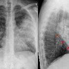
Bronchopneumonia is characterized by multiple small nodular or reticulonodular opacities which tend to be patchy and/or confluent. This represents areas of the lung where there are patches of inflammation separated by normal lung parenchyma. .
The distribution is often bilateral and asymmetric and predominantly involves the lung bases .
CT
Multiple foci of opacity can be seen in a lobular pattern, centered at centrilobular bronchioles. This may result in a tree-in-bud appearance. These foci of consolidation can overlap to create a larger heterogeneous confluent area of consolidation or 'patchwork quilt' appearance .
See also
Siehe auch:
und weiter:


 Assoziationen und Differentialdiagnosen zu Bronchopneumonie: