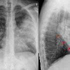
lobar pneumonia


Lobar pneumonia, also known as non-segmental pneumonia or focal non-segmental pneumonia , is a radiological pattern associated with homogeneous and fibrinosuppurative consolidation of one or more lobes of a lung in response to bacterial pneumonia.
The radiological appearance of lobar pneumonia is not specific to any single causative organism, although there are organisms which classically have a radiological presentation of lobar pneumonia. Streptococcus pneumoniae (also known as pneumococcus) is the most common causative organism of lobar pneumonia.
Epidemiology
Pneumonia is the most common cause of death due to infectious diseases in the United States, with an incidence of 11.6 per 1000 persons/year reported in one study . Incidence is higher at the extremes of age.
Clinical presentation
The presentation of lobar pneumonia depends on the severity of the disease, host factors and the presence of complications. Lobar pneumonia may present with a productive cough, dyspnea, pyrexia/fevers, rigours, malaise, pleuritic pain, and occasionally hemoptysis.
Key features on physical examination are dullness to percussion in a lobar pattern, bronchial breathing, and adventitious breath sounds. A pleural rub and reduced expansion on the affected side may be present .
Pathology
Consolidation in lobar pneumonia mainly affects the alveolar air spaces. There is characteristic relative sparing of the bronchi, creating the appearance of air bronchograms. The distribution of consolidation is lobar because of the spread of infection across segmental boundaries - facilitated by the pores of Kohn and the canals of Lambert - although limited by pleural boundaries.
The most common cause of lobar pneumonia is Streptococcus pneumoniae. Other causative organisms that may cause a lobar pattern include :
- Klebsiella pneumoniae
- Legionella pneumophila
- Haemophilus influenzae
- Mycobacterium tuberculosis
The gross and histologic appearance of the infected lung can be broken down into four stages of inflammation :
- congestion: hyperemia, with alveolar edema and bacterial proliferation
- red hepatisation: hemorrhagic inflammatory alveolar exudate
- grey hepatisation: fibrinopurulent inflammatory alveolar exudate
- resolution: final stage of processing the residual exudate
Red and grey hepatisation refers to the gross morphological appearance of a lung with inflammatory exudate in the alveolar spaces.
Radiographic features
Plain radiograph
Characteristically, there is homogeneous opacification in a lobar pattern. The opacification can be sharply defined at the fissures, although more commonly there is segmental consolidation . The non-opacified bronchus within a consolidated lobe will result in the appearance of air bronchograms. Strictly speaking, consolidation is not associated with volume loss; however, atelectasis can occur with small airway obstruction.
CT
Classically, lobar pneumonia appears as a focal dense opacification of the majority of an entire lobe with relative sparing of the large airways. There may be additional associated areas of ground-glass opacity in a lobar or segmental pattern, likely representing areas of partial involvement or simply atelectasis .
On contrast-enhanced CT, pneumonia often enhances less than atelectatic lung, although there is no clear Hounsfield unit threshold to distinguish the two. For example, one small study used a threshold of 85 HU to distinguish between atelectasis versus pneumonia on CT PE protocol with a sensitivity of 90% and specificity of 92% . However, there is overlap, and also factors such as pulmonary hemorrhage and underlying malignancy likely affect the lung density.
Treatment and prognosis
Radiological follow-up of lobar pneumonia is often recommended - one study found ~5% of initially suspected community-acquired pneumonia were re-diagnosed with malignant or important benign pulmonary pathology on follow-up chest radiographs/CT (average follow-up at 11.5 weeks) .
Complications
- pulmonary abscess
- pleural
- parapneumonic effusion - fibrinous inflammatory reaction to the adjacent pulmonary inflammation
- empyema - purulent fibrinous inflammatory reaction due to infectious spread into the pleural space
- note that both bland and purulent effusions may result in subsequent scarring/adhesions depending on the degree of fibroblastic organization
- disseminated infection
- bacteremia
- multiorgan infection
Differential diagnosis
For radiographic appearances of consolidation, consider other forms of lobar consolidation such as:
- atelectasis - tends to be associated with more volume loss, and is more enhancing compared to pneumonia
- pulmonary malignancy
- lung adenocarcinoma affecting an entire lobe
- forms of pulmonary lymphoma can affect an entire lobe
See also
Siehe auch:
und weiter:

 Assoziationen und Differentialdiagnosen zu Lobärpneumonie:


