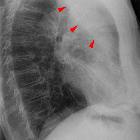
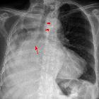
lobar lung collapse


Lobar collapse refers to the collapse of an entire lobe of the lung. As such it is a subtype of atelectasis (collapse is not entirely synonymous with atelectasis, which is a more generic term for 'incomplete expansion'). Individual lobes of the lung may collapse due to obstruction of the supplying bronchus.
Pathology
Most often collapse of most or all of a lobe is secondary to bronchial obstruction causing resorptive atelectasis.
Etiology
- luminal
- aspirated foreign material
- mucus plugging
- endobronchial mass
- misplaced endotracheal tube
- mural
- extrinsic
- compression by adjacent mass
Radiographic features
There are several classical rules that a lobar collapse follows :
- bowing or displacement of a fissure/s occurs towards the collapsing lobe
- a significant amount of volume loss is required to cause air space opacification
- the collapsed lobe is triangular or pyramidal in shape, with the apex pointing to the hilum
- the collapsed lung peripherally maintains contact with the costal parietal pleura, except:
- in RML collapse where the lobe collapses adjacent to the mediastinum
- in the presence of pleural effusion
- in the presence of pneumothorax
Several factors may influence the typical appearance of lobar collapse, including pre-existing lung disease, amount of volume loss, concomitant consolidation, pleural effusion or the presence of pneumothorax.
Plain radiograph
Generally, there is pulmonary air space opacification but the appearance on chest x-ray varies according to the lobe involved and are discussed separately:
- right upper lobe collapse
- right middle lobe collapse
- right lower lobe collapse
- left upper lobe collapse
- left lower lobe collapse
Some features, however, are generic markers of volume loss and are helpful in directing one's attention to the collapse, as well as enabling distinction from opacification of the lobe without collapse (i.e. consolidation e.g. lobar pneumonia). These features include :
- direct signs
- displacement of fissures
- crowding of pulmonary vessels
- indirect signs
- elevation of the ipsilateral hemidiaphragm
- crowding of the ipsilateral ribs
- shift of the mediastinum towards the side of atelectasis
- compensatory hyperinflation of normal lobes
- hilar displacement towards the collapse
- shifting granuloma sign
CT
Lobar collapse is usually trivially easy to identify on CT, but identification of the cause is not always easy, as the collapsed lung can make identification of an obstructing lesion difficult. The density of the collapsed lobe is high post contrast administration.
Siehe auch:
- Atelektase
- Mittellappenatelektase
- Lungenkarzinom
- left lower lobe collapse
- right upper lobe collapse
- left upper lobe collapse
- Unterlappenatelektase rechts
und weiter:

 Assoziationen und Differentialdiagnosen zu lobar collapse:


