right lower lobe collapse


A right lower lobe (RLL) collapse has distinctive features, and is usually relatively easily identified. The absence of overlying cardiomediastinal outline makes it easier to appreciate than left lower lobe collapse.
Findings of lower lobe collapse can be grouped together as they are almost identical on both sides.
For a general discussion please refer to the article on lobar collapse.
Radiographic features
Plain radiograph
Complete collapse of the right lower lobe, although usually easily seen when looked for can be missed if there is very little consolidation and film is rotated. Features include :
- triangular opacity at the right lower zone (usually medially) with the apex pointing towards the right hilum
- obscuration of the medial aspect of the dome of right hemidiaphragm
- inferior displacement of the right hilum
- descending interlobar pulmonary artery is not visible
- preservation of a clear right heart border, which is contacted by the right middle lobe (cf. obscuration seen in RML collapse or consolidation)
- inferior displacement of the horizontal fissure
Non-specific signs indicating right sided atelectasis may also be present.
They include:
- elevation of the right hemidiaphragm
- crowding of the right sided ribs
- shift of the mediastinum to the right
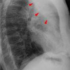
On the lateral projection the findings are usually obvious:
- triangular opacification in the lower posterior chest
- right hemidiaphragm is obscured posteriorly and the lower thoracic vertebrae appear denser than normal (they are usually more radiolucent than the upper vertebrae)
- oblique fissure displaced posteroinferiorly but may be invisible with severe collapse of the RLL as it fissure rotates posteromedially becoming non-tangential to the x-ray beam
- inferior displacement of the right hilum
If there is an obstructing lesion in the bronchus intermedius, there will be signs of both RML and RLL collapse.
CT
- triangular opacification in axial images, thinner at the hilum, against the posterior mediastinum and medial hemidiaphragm
- oblique fissure rotates posteromedially or posteriorly
- compensatory hyperinflation of the right upper lobe
Differential diagnosis
The characteristic shape associated with volume loss usually does not allow for any significant differential diagnosis. As always one should consider:
- consolidation (of the medial basal segment of the right lower lobe) which will show absence of signs of volume loss
- pulmonary or posterior mediastinal mass
This location is also common for pulmonary sequestration.
Related articles
Siehe auch:
- Atelektase
- Mittellappenatelektase
- Lungensequester
- left lower lobe collapse
- left upper lobe collapse
- lobar collapse
- Normale Herzkonfiguration im Röntgen-Thorax
- rechter Unterlappen
und weiter:


 Assoziationen und Differentialdiagnosen zu Unterlappenatelektase rechts: