normal contours of the cardiomediastinum on chest radiography

Normal
contours of the cardiomediastinum on chest radiography • Cardiomediastinal anatomy on chest radiography (annotated images) - Ganzer Fall bei Radiopaedia

Normal
contours of the cardiomediastinum on chest radiography • Cardiomediastinal outlines on chest x-ray - Ganzer Fall bei Radiopaedia
Cardiac
valves • Cardiomediastinal anatomy on chest radiography (annotated images) - Ganzer Fall bei Radiopaedia
A detailed understanding of the structures that make up the normal contours of the heart and mediastinum (cardiomediastinal contour) on chest radiography is essential if abnormalities are to be detected.
Frontal view (PA/AP)
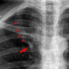
Right cardiomediastinal contour
From superior to inferior:
- right paratracheal stripe
- seen in two thirds of normal films
- made up of right brachiocephalic vein and SVC
- arch of the azygous vein
- ascending aorta in older individuals often projects to the right of the SVC
- superior vena cava (SVC)
- right atrium
- inferior vena cava (IVC)
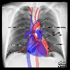
Left cardiomediastinal contour
From superior to inferior:
- left paratracheal stripe
- made up of left common carotid artery, left subclavian artery and the left jugular vein
- aortic arch +/- aortic nipple (left superior intercostal vein)
- pulmonary trunk
- auricle of left atrium
- left ventricle
Lateral view
Anterior cardiomediastinal contour
From superior to inferior:
- superior mediastinum
- great vessels
- thymus
- ascending aorta
- right ventricular outflow tract
- right ventricle
Posterior cardiomediastinal contour
From superior to inferior:
- left atrium and pulmonary veins
- left ventricle
- inferior vena cava
Siehe auch:
- Herzkonfiguration
- Thymus
- Vena cava inferior
- Vena azygos
- left ventricular enlargement
- right paratracheal stripe
- Aortenbogen
- vergößerter linker Vorhof
- Vena cava superior
- cardiac chamber enlargement
- Vergrößerung rechter Vorhof
- Vergrößerung rechter Ventrikel
und weiter:
- Mittellappenatelektase
- Röntgen-Thorax
- left lower lobe collapse
- Tumoren des vorderen oberen Mediastinums
- right upper lobe collapse
- Herzfehler
- acyanotic congenital heart disease
- left upper lobe collapse
- enlargement of the cardiac silhouette
- Varianten der Herzanatomie
- Unterlappenatelektase rechts
- CXR approach to congenital heart disease
- posterior mediastinal mass in the exam
- chest x-ray appeoach to congenital heart disease
- congenital heart disease - chest x-ray approach
- Thorax Onlinekurs
- atrial escape
- Zyanotischer Herzfehler
- right upper lobe collapse in the exam
- left upper lobe collapse in the exam
- Aortentrauma


 Assoziationen und Differentialdiagnosen zu Normale Herzkonfiguration im Röntgen-Thorax: