superior vena cava (SVC)

Common
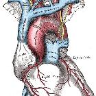
carotid artery • Great vessels (Gray's illustration) - Ganzer Fall bei Radiopaedia


Superior vena
cava • Superior vena cava anatomy - Ganzer Fall bei Radiopaedia

Inferior vena
cava • Truncal venous development (Gray's illustrations) - Ganzer Fall bei Radiopaedia

Inferior vena
cava • Caval variants (illustrations) - Ganzer Fall bei Radiopaedia
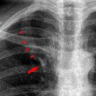
The superior vena cava (SVC) is a large valveless venous channel formed by the union of the brachiocephalic veins. It receives blood from the upper half of the body (except the heart) and returns it to the right atrium.
Gross anatomy
The superior vena cava begins behind the lower border of the first right costal cartilage and descends vertically behind the first and second intercostal spaces to drain into the right atrium at the superior cavoatrial junction (at the level of the third costal cartilage). Its lower half is covered by the fibrous pericardium, which it pierces at the level of the second costal cartilage .
Tributaries
- azygos vein
- small veins draining the pericardium and other mediastinal structures
Relations
- left lateral: aortic arch, trachea
- right lateral: pleura, right upper lobe, right phrenic nerve
- anteriorly: thymus, manubrium
- posteriorly: azygos vein
- superiorly: brachiocephalic veins, superior thoracic aperture
- inferiorly: pericardium, right atrium
Variant anatomy
- brachiocephalic veins: drain into the right atrium separately
- SVC duplication
- left sided SVC
Related pathology
Siehe auch:
und weiter:

 Assoziationen und Differentialdiagnosen zu Vena cava superior:


