Azygoskontinuität


Azygos continuation of the inferior vena cava (also known as the absence of the hepatic segment of the IVC with azygos continuation) is an uncommon vascular anomaly and is a cause of a dilated azygos vein.
Terminology
Spelling it "azygous" when referring to the vein is incorrect, regardless of whether British or American English . Azygous is a word in English meaning 'without pair', but in the context of anatomy, see Terminologia Anatomica, azygos vein is the sole correct spelling.
Epidemiology
Azygos continuation of the IVC has a prevalence ~1.5% (range 0.2-3%) .
Clinical presentation
In most cases, it is found incidentally in asymptomatic patients. An association with congenital heart disease and asplenia or polysplenia syndromes have been reported.
It is important to recognize the enlarged azygos vein at the confluence with the superior vena cava and in the retrocrural space to avoid misdiagnosis as a right-sided paratracheal mass or retrocrural adenopathy.
Preoperative knowledge of anatomy may be important in planning cardiopulmonary bypass and to avoid difficulties in catheterizing the heart.
Gross anatomy
Contrary to the name, the post-hepatic segment of the IVC (derived from the right vitelline vein) is present, draining only the hepatic veins into the right atrium. Instead, there is an interruption of the infrahepatic/suprarenal portion of the IVC. This represents a failure of fusion of the vitelline and subcardinal embryological portions of the IVC.
The renal segment of the IVC, therefore, drains via the azygos vein (or in rare cases, the hemiazygos vein if there is a left-sided IVC).
Associations
Traditionally it was thought to be strongly associated with other congenital heart defects and polysplenia. However, the widespread use of cross-sectional imaging has revealed that azygos IVC continuation can occur in isolation and can be asymptomatic.
A commonly associated abnormality is the duplication of inferior vena cava (IVC). In these cases, usually, the left IVC typically ends at the left renal vein which crosses anterior to the aorta to join the right IVC. Then the right IVC remains as the azygos vein, missing the liver. The azygos vein joins the superior vena cava (SVC) at the normal location in the right paratracheal space.
Other arrangements in the duplicated inferior vena cava drainage can be found, such as:
- double IVC with retroaortic right renal vein and hemiazygos continuation of the IVC
- double IVC with azygos and hemiazygos continuation of each duplicated IVC
Radiographic features
Plain radiograph
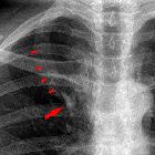
On frontal chest radiograph, the mediastinum is widened and the azygos arch enlarged.
CT and MRI
Cross-sectional imaging usually has no difficulty in identifying the abnormality, demonstrating the anatomy elegantly (see above).
Treatment and prognosis
As an isolated finding, azygos continuation of the IVC requires no treatment.
Preoperative knowledge of anatomy may be important in planning cardiopulmonary bypass and to avoid difficulties in catheterizing the heart.
Siehe auch:
- Duplikatur der Vena cava inferior
- Varianten der Vena cava inferior
- Situs inversus
- Vena azygos
- Varianten der Vena azygos
- erweiterte Vena azygos
- Vena cava superior
- Lebervenen
- Polysplenie-Syndrom
- Agenesie Vena cava inferior
und weiter:


 Assoziationen und Differentialdiagnosen zu Azygoskontinuität: