duplication of inferior vena cava


Duplication of the inferior vena cava is a relatively rare vascular anomaly, but this caval abnormality needs to be recognized, especially in association with renal anomalies like crossed fused ectopia or circumaortic renal collar .
Epidemiology
The incidence of inferior vena cava duplication is reported to be ~1.5% (range 0.2-3%) .
Associations
- horseshoe kidney
- circumaortic renal collar
- crossed fused ectopia
- retroaortic left renal vein
- cloacal exstrophy
Case reports of association with ureteropelvic junction anomaly and retrocaval ureters have been described.
Clinical presentation
Usually, an incidental detection, while evaluating genitourinary anomalies.
Pathology
The inferior vena cava has a convoluted development during the 7-10weeks of gestation .
- posterior cardinal vein appears first but forms only the distal IVC i.e. iliac bifurcation.
- subcardinal veins (2) appear next, left subcardinal vein regresses, and right subcardinal vein forms the suprarenal IVC.
- supracardinal veins (2) appear last, left supracardinal vein regresses, and right supracardinal vein forms infrarenal IVC.
IVC duplication results from a persistent left supracardinal vein.
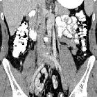
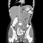
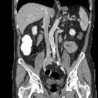
Radiographic features
Duplicated left sided IVC is usually seen as a continuation of the left common iliac vein, crossing anterior to the aorta at the level of renal vein to join the right sided IVC.
Differential diagnosis
An important differential is transposition of IVC. The renal vein is an important landmark for this differential. IVC continues on both sides of the aorta, in duplicated IVC. However, in transposition of the IVC, it continues on the left side of the aorta only.
Siehe auch:
- persistierende linke obere Hohlvene
- Varianten der Vena cava inferior
- Azygoskontinuität
- Hufeisenniere
- gekreuzte Nierendystopie mit Verschmelzung
- Kloakenekstrophie
- circumaortic renal collar
- Varianten der Vena cava
- linksseitige Vena cava inferior
- right double inferior vena cava
- caval abnormality
und weiter:


 Assoziationen und Differentialdiagnosen zu Duplikatur der Vena cava inferior:


