Situs ambiguus


Heterotaxy syndrome or situs ambiguus (also commonly, but etymologically less correctly, spelled situs ambiguous) is a disturbance in the usual left-right distribution of the thoracic and abdominal organs that does not entirely correspond to the complete mirror image (situs inversus).
It occurs from an early embryological developmental disturbance with most cases being sporadic. It is also classified under the group of cardiosplenic syndromes.
Terminology
Specialists employ varying definitions of heterotaxy syndrome. In this article, the term heterotaxy syndrome is used synonymously with situs ambiguus . Some authors instead refer to heterotaxy syndrome as a specific subset of situs ambiguus, such as that with the presence of complex congenital heart defects like cardiac isomerism .
Epidemiology
The true incidence is not known, but some sources have estimated it to be around 1 per 8,000-25,000 live births. Approximately 20-25% are associated with the immotile cilia syndrome (including Kartagener syndrome).
Clinical presentation
This is dependent on severity of the isomerism and presence of associated abnormalities. Cyanotic congenital heart disease is the main presentation in right isomerism. Left isomerism tends to present later in childhood or even in adulthood since it is less associated with complex congenital heart disease. Intestinal malrotation with midgut volvulus may also be a presenting feature.
Pathology
Bronchial anatomy as a key
Bronchial anatomy accurately reflects atrial situs. The bronchial anatomy on the left and right can be recognized on a well-penetrated radiograph and consists of two main bronchi that are anatomically different:
- hyparterial bronchus (below artery): supplies the bilobed left lung
- eparterial bronchus (alongside the artery): supplies the trilobed right lung
In situs ambiguus, there is duplication of either the hyparterial or eparterial bronchus. The associated atria are also duplicated and there are specific changes below the diaphragm (although not left-sided or right-sided duplication)
Radiographic features
Imaging features can be extremely complex.
There is duplication of the left or right-sided intrathoracic contents with associated changes below the diaphragm. Classically, there is malposition of the liver, stomach, and spleen (which may be absent). Additionally, the vascular supply above and below the diaphragm may be altered significantly (including SVC duplication) .
Left isomerism
Known as polysplenia syndrome:
- multiple splenules without a parent spleen
- azygos or hemiazygos continuation of the inferior vena cava
- bilateral hyparterial bronchi
- bilateral bilobed lungs
- bilateral pulmonary/left atria
- midline/transverse liver
- intestinal malrotation
Right isomerism
Known as asplenia syndrome:
- severe cyanotic congenital heart diseases
- absence of spleen
- bilateral eparterial bronchi
- bilateral trilobed lungs
- bilateral right atria
- midline/transverse liver
- intestinal malrotation
Imaging workup
Assessment of the intrathoracic contents can be made with plain film, echocardiography, CT, and MRI as well as angiography. Below the diaphragm, the abdominal contents can be imaged with ultrasound, GI contrast studies, CT and MRI.
A minimal workup should include :
- chest radiograph
- echocardiogram: to assess for congenital heart disease
- abdominal ultrasound: to assess intra-abdominal contents (especially spleen)
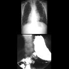
- upper GI series: to rule out malrotation
Treatment and prognosis
Treatment is completely dependent on the malformations (that occur to varying degrees) and the impact that they have clinically.
Specific treatment of congenital heart disease can be seen in their separate articles.
See also
Siehe auch:
- persistierende linke obere Hohlvene
- Azygoskontinuität
- Situs inversus
- intestinale Malrotation
- Herzfehler
- Kartagener-Syndrom
- primäre ciliäre Dyskinesie
- totale Lungenvenenfehlmündung
- situs classification
- Polysplenie-Syndrom
- Heterotaxie
und weiter:


 Assoziationen und Differentialdiagnosen zu Situs ambiguus:


