Lungenvenenfehlmündung

Fehllage ZVK
V. juguluaris interna links bei partieller Fehlmündung der Lungenvenen (PAPVR) des linken Oberlappens in die V. brachiocephalica sinistra. Bettlunge mit Fehllage des ZVKs und CT coronare MIP ohne ZVK zur Darstellung der Anatomie. (Bildqualität bei Adipositas permagna eingeschränkt.)


Lungenvenenfehleinmündung
linker Oberlappen in V. brachiocephalica sinistra in der Computertomografie: Links 3 Schichten axial (mitabgebildete axilläre Lymphadenopathie), rechts Volumen Rendering und parakoronar.

Lungenvenenfehlmündung
vom linken Oberlappen in die V. brachiocephalica sinistra: Die Lungenvene des Oberlappens links verläuft vom Hilus kommend links neben dem Aortenbogen schräg nach oben, wo sich das Blut mit dem stark kontrastierten Blut aus der V. subclavia bei Kontrastmittelgabe über den linken Arm vermischt.

Anomalous
pulmonary venous drainage: a pictorial essay with a CT focus. Scimitar Syndrome. a Coronal oblique maximum intensity projection (MIP) CT images. A large right sided pulmonary vein is seen supradiaphragmatic inferior vena cava (IVC) in a patient with Scimitar Syndrome. b Axial CT image of same patient as in (a). The white arrow indicates the anomalous right pulmonary vein. The asymmetry of the pulmonary vasculature provides a clue to identifying the abnormality. The right hemidiaphragm is elevated consistent with hypoplasia of the right lower lobe, a common association with anomalous pulmonary venous drainage. c Chest radiograph of a paediatric patient, demonstrating a tubular opacity in the right paracardiac region consistent with a scimitar vein (arrow). d Coronal MIP CT image showing a large right pulmonary vein draining into the infradiaphragmatic IVC


Fehllage ZVK
V. juguluaris interna links bei partieller Fehlmündung der Lungenvenen (PAPVR) des linken Oberlappens. In der Blutgasanalyse aus dem ZVK ergab sich weder eine typisch arterielle noch eine typisch venöse Sauerstoffsättigung. Bettlunge mit Fehllage des ZVKs. Darunter coronare MIP. Je 3 Schichten axial und coronar zum Verlauf der Vene, die in die V. brachiocephalica sinistra mündet. (Bildqualität bei Adipositas permagna eingeschränkt.)

Imaging of
congenital lung diseases presenting in the adulthood: a pictorial review. Different examples of partial anomalous pulmonary venous return (PAPVR). Axial CT (a) and 3D volume-rendered (b) images demonstrate anomalous right upper lobe pulmonary veins draining into the superior vena cava (arrows). The anomalous left upper lobe pulmonary vein draining into the brachiocephalic vein is observed on coronal CT (c) and 3D volume-rendered (d) images (arrows). An anomalous pulmonary vein is seen on coronal CT image (e, arrow) and central venous catheter malposition with the catheter tip extending towards the right lung in the same patient observed on chest X-ray (f, arrow)

Imaging of
congenital lung diseases presenting in the adulthood: a pictorial review. Mixed lesions-Kartagener syndrome and partial anomalous pulmonary venous return (PAPVR) in a 20-year-old man. Axial (a) and coronal (c) CT images show anomalous right upper lobe pulmonary veins draining into the right brachiocephalic vein. Bronchiectasis, bronchial wall thickening, and centrilobular opacities predominantly affecting lower lobes of the lungs are seen on axial CT image (b). Coronal CT image (c) demonstrates both PAPVR (arrow) and bronchiectasis. 3D volume-rendered reconstruction image (d) shows PAPVR (arrow)

Anomalous
pulmonary venous drainage: a pictorial essay with a CT focus. Embryology of anomalous pulmonary venous drainage. This diagram illustrates right sided pulmonary veins failing to establish a normal connection with the left atrium and instead draining into the inferior vena cava. UV = Umbilicovitelline vein; CCV = Common cardinal vein; LB = Lung buds; LA = Left atrium; RA = Right atrium; LV = Left ventricle; RV = Right ventricle; SVC = Superior vena cava; IVC = Inferior vena cava; ARPV = Anomalous right pulmonary vein

Anomalous
pulmonary venous drainage: a pictorial essay with a CT focus. Different variations of anomalous right superior pulmonary vein. (a) Axial image at the level of the main pulmonary artery. Right superior pulmonary vein (RSPV) draining into the superior vena cava (SVC) only (arrow). No sinus venosus defect was demonstrated. (b) Axial oblique CT. RSPV with a connection to the superior vena cava/right atrium (SVC/RA) confluence (arrow), but then continuing to drain into the left atrium (arrowhead). No direct sinus venosus defect. The right ventricle is dilated. (c) Axial oblique CT. RSPV draining into the SVC/RA confluence (white arrow) with a superior sinus venosus defect (black arrowhead) and right ventricular dilatation. (d) Axial oblique CT image. Anomalous RSPV is seen draining into the SVC (white arrow). Superior sinus venosus defect is demonstrated (black arrow) as well as an atrial secondum defect (black arrowhead)

Anomalous
pulmonary venous drainage: a pictorial essay with a CT focus. Anomalous drainage into the azygos vein. Images a and b are from an adolescent patient with anomalous pulmonary venous drainage into the azygos vein. Images c and d are from a paediatric patient with anomalous pulmonary venous drainage identified on echocardiography. a Sagittal maximum intensity projection (MIP) CT image showing the right superior pulmonary vein (white arrow) draining into a dilated azygos vein (labelled AZ). b Three-dimensional (3D) CT reconstruction of patient in (a), posterior view. The black arrow indicates the right superior pulmonary vein draining into the azygos vein. c 3D CT reconstruction. Most of the right sided pulmonary veins are seen draining into a dilated azygos vein (labelled AZ). d Axial CT image of the patient described in (c). Only a very small right middle pulmonary vein is seen draining normally into the left atrium (white arrow). Note the dilated right atrium and right ventricle consistent with a significant left-to-right shunt

Anomalous
pulmonary venous drainage: a pictorial essay with a CT focus. Anomalous left superior pulmonary vein. a Axial oblique maximum intensity projection (MIP) CT image. Anomalous left superior pulmonary vein (LSPV) drains into the left brachiocephalic vein (LBCV) (white arrow). AO – Aorta. b Three-dimensional CT reconstruction demonstrating the anomalous LSPV draining into the LBCV (white arrow)

Anomalous
pulmonary venous drainage: a pictorial essay with a CT focus. Anomalous left superior pulmonary vein (LSPV) may mimic a left sided superior vena cava (SVC) on axial CT images. Patient 1. Bilateral SVCs with anomalous left SVC draining into the left atrium (a and b). a Bilateral SVCs. Right SVC (arrowhead) and small left sided SVC (white arrow). b Coronal MIP CT in the same patient showing the anomalous left SVC draining into the left atrium (white arrow). Normal right-sided SVC (thin white arrow). Patient 2. Partial anomalous pulmonary venous drainage (c and d). c Anomalous LSPV (white arrow), which on single axial slice is similar in appearance to the left-sided SVC in (a). d Coronal MIP CT showing the anomalous LSPV (white arrow) draining into the left brachiocephalic vein (LBCV). Normal right SVC (thin white arrow)

Anomalous
pulmonary venous drainage: a pictorial essay with a CT focus. Anomalous right-sided pulmonary venous drainage with a superior sinus venosus defect. a Axial oblique CT MPR images show an anomalous right superior pulmonary vein (RSPV) draining into the superior vena cava/right atrium (SVC/RA) junction (white arrow) with an associated superior sinus venosus defect (SSVD, arrowhead) connecting the left atrium (LA) and right atrium (RA). b Post surgical repair the right superior pulmonary vein is now directed into the left atrium with closure of the SSVD. c Sagittal oblique CT images showing the SVC overriding a SSVD between the two atria. d Post surgery the SSVD has been repaired, the SVC (black arrow) drains directly into the right atrium

School ager
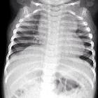
with dyspneaCXR AP (above) shows a hypoplastic right lung, AP arterial (left) and venous (right) phases of a pulmonary artery angiogram show a right pulmonary (scimitar) vein draining into the inferior vena cava.The diagnosis was partial anomalous pulmonary venous return.

Anomalous
pulmonary venous drainage: a pictorial essay with a CT focus. Anomalous right inferior pulmonary vein drain. Adult patient presenting with breathlessness. a Three-dimensional (3D) CT reconstruction. A large right inferior pulmonary vein is seen draining most of the right upper lobe into the supradiaphragmatic inferior vena cava (arrow). Most of the lower lobe is seen draining normally into the left atrium (arrowhead). b Sagittal MIP CT image shows a hypoplastic right lower lobe (arrow – oblique fissure). The large curvilinear right pulmonary vein is seen draining the right upper lobe

Anomalous
pulmonary venous drainage: a pictorial essay with a CT focus. Chest radiograph findings in anomalous pulmonary venous drainage- Focal mediastinal widening to the left of the aortic knob. a Patient 1. Postero-anterior (PA) chest radiograph (CXR). Vascular shadow adjacent to the aortic knuckle corresponds to anomalous left superior pulmonary vein (LSPV) in (b). b Coronal maximum intensity projections (MIP) CT image demonstrating anomalous LSPV (black arrow) draining into the brachiocephalic vein (LBCV). This is the most common left sided partial anomalous pulmonary venous drainage pattern. c Patient 2. Posteroanterior chest radiography. The LSPV can be seen as a prominent vascular shadow lateral to the aortic knuckle and left subclavian artery (black arrow). d Coronal MIP MR angiography demonstrating LSPV (arrow) draining into the left BCV (LBCV)

Anomalous
pulmonary venous drainage: a pictorial essay with a CT focus. Effects of a longstanding left-to-right shunt in patients with PAPVD. a Right superior pulmonary vein (RSPV) draining into the superior vena cava (SVC). Right sided pulmonary plethora is seen. b Right atrial dilatation, cardiomegaly and features of pulmonary oedema. Subsequent computed tomographic pulmonary angiography (CTPA) demonstrated a RSPV draining into the SVC with dilated right heart chambers. c Patient with anomalous right superior and middle pulmonary veins draining into the superior vena cava/right atrium confluence. A chest radiograph demonstrates cardiomegaly and central pulmonary artery dilatation secondary to long-term left-to-right shunt

Anomalous
pulmonary venous drainage: a pictorial essay with a CT focus. Total anomalous pulmonary venous drainage (TAPVD) repair. a Three-dimensional (3D) CT reconstruction. An adult patient presenting with breathlessness had a computed tomographic pulmonary angiogram (CTPA). A single left pulmonary vein (LPV) crosses the midline to form a single confluence with the right pulmonary veins (RPV) and joins the left atrium (LA) via a baffle (white arrow). A residual anomalous left superior pulmonary vein (white arrowhead) is seen draining into the left brachiocephalic vein (LBCV); presumed previous type 4 TAPVD. Incidentally the pulmonary vessels in the right lower lobe were tortuous and dilated with a suspected arteriovenous malformation. b Three dimensional (3D) CT reconstruction. A paediatric patient with a Type 2 TAPVD repair. The pulmonary veins (white arrowheads) can be seen forming a single venous confluence posterior to the LA. This has been reconnected to the LA via a small baffle (white arrow). c Sagittal CT image. Type 2 TAPVD. A single venous confluence of the pulmonary veins is present (asterisk) posterior to the left atrium (LA) with a focal narrowing where this has been surgically connected to the LA (black arrow). d Axial CT image. This again shows the posterior venous confluence of the pulmonary veins (black arrow)


Anomalous
pulmonary venous drainage: a pictorial essay with a CT focus. Anomalous right superior and middle pulmonary veins. (a) Coronal oblique maximum intensity projection CT image. The right superior pulmonary vein is seen draining into the superior vena cava (arrow). Just inferior to this the right superior and inferior middle pulmonary veins are also seen draining separately into the superior vena cava/right atrium confluence (arrowheads). b Axial oblique MIP CT 5-chamber view, demonstrating a superior sinus venosus defect (black arrow), anomalous middle pulmonary vein (white arrowhead) and marked right ventricular dilatation secondary to the left-to-right shunt
 nicht verwechseln mit: meandernde Pulmonalvenen
Lungenvenenfehlmündung
Während bei der partiellen Lungenvenenfehlmündung nur ein Teil der Lungenvenen falsch, also nicht in den linken Vorhof mündet, sind dies bei der totalen Lungenvenenfehlmündung alle Lungenvenen. Diese Konstellation gehört somit zu den zyanotischen Herzfehlern und ist unmittelbar nach der Geburt eine entsprechend bedrohliche Situation. Dagegen wird die partielle Lungenvenenfehlmündung je nach Anteil des falsch zurückgeführten Blutes manchmal erst später, bisweilen nur zufällig entdeckt.
Die bekannteste Lungenvenenfehlmündung ist das Scimitar-Syndrom.
Siehe auch:
- Lungensequester
- Atriumseptumdefekt
- Lungenhypoplasie
- Scimitar-Syndrom
- Situs ambiguus
- Snowman heart
- totale Lungenvenenfehlmündung
- partielle Lungenvenenfehlmündung
- pulmonary venolobar syndrome
- Zyanotischer Herzfehler
- meandernde Pulmonalvenen
und weiter:

 Assoziationen und Differentialdiagnosen zu Lungenvenenfehlmündung:


