Vena cava inferior Thrombose


 nicht verwechseln mit: Scheinthrombose Vena cava inferior
Inferior vena caval thrombosis is an essential diagnosis while evaluating any neoplastic lesion, or portal hypertension. It is also important to differentiate bland thrombus from tumor thrombus.
Clinical features
A patient can present with many features which include
- bilateral pedal edema
- Budd-Chiari syndrome
- pulmonary embolism
Pathology
Classification
Inferior vena cava thrombus can be classified under two broad headings:
Bland thrombus
Bland thrombus can be an isolated thrombus or commonly arising from deep vein thrombosis of the lower extremities.
Etiology
- hypercoagulable states
- venous stasis
- compression by neoplastic lesions, lymph nodes, retroperitoneal masses/fibrosis or hemorrhage
- foreign bodies: vena cava filter, catheters
- extension from benign tumors: angiomyolipoma, inferior vena cava leiomyoma, adrenal pheochromocytoma
- traumatic liver injury: very rare
Tumor thrombus
While any neoplastic lesions can cause inferior vena cava thrombosis, renal cell carcinomas are the most common malignancy to extend into the inferior vena cava and this has important implications for surgical management .
Other tumors that have a tendency to cause inferior vena cava thrombosis are hepatocellular carcinoma, adrenocortical cancer, and Wilms tumor, primary leiomyoma or leiomyosarcoma of the inferior vena cava.
According to TNM staging of renal cell carcinoma, tumor spread into the infra diaphragmatic inferior vena cava is the T3c stage, while extension into the supradiaphragmatic inferior vena cava is T4b stage. Also, subclassification into infrahepatic, hepatic, and suprahepatic extension can further help the surgeon.
Radiographic features
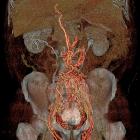
On all imaging, they appear as persistent filling-defects within the inferior vena cava. Chronic thrombosis can lead to pericaval and periaortic collateral formation .
Differentiating bland and tumor thrombus
- a bland thrombus result from external compression of the inferior vena cava by a neoplastic lesion (i.e. no direct invasion), so the inferior vena cava is usually narrowed at the site of thrombosis; in contrast, tumor thrombus expands the inferior vena cava
- tumor thrombus will often show continuity with primary tumor
- in arterial phase, neovascularity may be appreciated in tumor thrombus
Differential diagnosis
Conditions that may mimic thrombus include:
- pseudothrombosis caused by laminar flow of enhanced blood from renal veins streaming parallel to the column of unopacified blood returning from the lower body; delayed images will show resolution of filling defect
- pseudolipoma (paracaval lipoma) is the partial volume artifact due to pericaval fat above caudate lobe, seen commonly in patients with chronic liver disease; coronal reformats usually clear the doubt
Siehe auch:
- Azygoskontinuität
- Vena cava inferior
- lipomatöse Impression der Vena cava inferior
- erweiterte Vena azygos
- Tumorthrombus
- Thrombose Vena cava superior
- Leiomyosarkom der Vena cava inferior
- Thrombose der Vena cava
- Thrombose der Vena cava inferior beim Neugeborenen
- iliofemorale tiefe Venenthrombose
- Obstruktion Vena cava inferior
- Inferior vena caval thrombosis secondary to supra-renal inferior vena caval atresia or agensis (IVCA)
- Scheinthrombose Vena cava inferior
- laminarer Fluss in der Vena cava inferior mit Scheinthrombose
und weiter:


 Assoziationen und Differentialdiagnosen zu Vena cava inferior Thrombose: