superior vena cava obstruction
Superior vena cava (SVC) obstruction can occur from extrinsic compression, intrinsic stenosis, or thrombosis. Malignancies are the main cause and are considered an oncologic emergency. Superior vena cava syndrome (SVCS) refers to the clinical syndrome with symptoms that results from this obstruction.
Clinical presentation
Clinical presentation depends on the speed, severity, and location of superior vena cava obstruction . Collateral drainage may develop with slow obstruction and patients may have no or only mild symptoms.
With acute superior vena cava obstruction, symptoms include facial and neck swelling, facial flushing, bilateral upper extremity swelling, neurological signs, dyspnea, headache, and cough.
The Pemberton maneuver might be a useful physical examination tool. The maneuver is achieved by having the patient elevate both arms until they touch the sides of the face. A positive Pemberton's sign is marked by the presence of facial congestion and cyanosis, as well as respiratory distress after approximately one minute.
Pathology
Etiology
- malignancy (~90% of cases), most commonly :
- central venous catheters
- pacemaker wires
- fibrosing mediastinitis
- luetic aneurysm
- Behcet disease
Pathophysiology
In long-standing cases with 60% or more stenosis, collateral channels are formed to restore venous return. Various collaterals are formed depending on the site of the obstruction:
- pre azygos: in these conditions mainly the right superior intercostal veins serve as the collateral pathway to drain into the azygos vein.
- azygos: when the azygos vein is also obstructed the collateral circulation establishes between SVC and IVC via minor communicating channels i.e. internal mammary veins, superior and inferior epigastric veins to iliac veins, and finally into the IVC.
- post azygos: in this case, the blood from the SVC is distributed into the azygos and hemiazygos and then into the IVC tributaries i.e. ascending lumbar and lumbar veins.
The most efficient collateral system is right superior intercostal and azygos circulation. For this reason, most of the patients with pre azygos obstruction of SVC remain asymptomatic for a long period of time.
Radiographic features
Plain radiograph
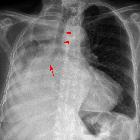
Indirect signs on chest x-ray, such as superior mediastinal widening and right hilar prominence that may indicate the presence of mediastinal mass. Aortic nipple sign is also a feature.
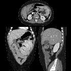
CT
Is the imaging modality of choice. Enhanced CT shows the location and severity of the SVC obstruction, superimposed thrombosis, a mediastinal mass or lymphadenopathy, collateral vessels, and associated lung masses.
See: superior vena cava obstruction: grading
Treatment and prognosis
Treatment of SVCS will depend on the cause of the compression. Thrombolysis and anticoagulation may be indicated for thrombosis. In cases of compression, endovascular treatment with self-expandable bare stents is an effective SVCS therapy . With carcinoma or infection, specific drugs or radiation may be used .
Siehe auch:
- Rippenusuren
- Lungenarterienembolie
- Lungenkarzinom
- Spannungspneumothorax
- Vena cava superior
- fibrosierende Mediastinitis
- Burkitt-Lymphom
- Asthma bronchiale
- Stenose Vena cava superior bei liegendem Port
- differential diagnosis of dilated mammary veins
- acute leukemia
- Perikardtamponade
- Herzklappeninsuffizienz
- lymphoblastic lymphoma
- pre-T-cell lineage acute lymphoblastic leukemia
- Linksherzschwäche
- Downhill Ösophagusvarizen
und weiter:


 Assoziationen und Differentialdiagnosen zu Obere Einflussstauung: