aortic arch


The aortic arch represents the direct continuation of the ascending aorta and represents a key area for a review of normal variant anatomy and a wide range of pathological processes that range from congenital anomalies to traumatic injury.
Summary
- origin: continuation of the ascending aorta at the level of the sternomanubrial joint, in the plane of Ludwig
- course: an arch from right to left and front to back; lies anterior and to the left of the trachea
- main branches: brachiocephalic trunk, left common carotid artery and left subclavian artery.
- supply: head, neck and upper limbs
- termination: adjacent to the lower border of T4 in the plane of Ludwig where it continues as the descending aorta
- key relationships: trachea, pulmonary trunk and arteries
Gross anatomy
Origin
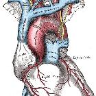
The aortic arch represents the continuation of the ascending aorta and is nominally defined as starting at the level of the transthoracic plane of Ludwig, a horizontal plane from the sternomanubrial angle to the T4 vertebral body. The sternomanubrial joint is the same level as the second sternocostal articulation.
Course
It courses in a narrow arch from ventral to dorsal and from right to left such that at the end of the arch it sits to the left of midline, adjacent to the thoracic vertebral column. Its peak is at the T3/4 level.
Branches
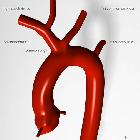
Three main branches originate from the upward convexity of the arch in the majority (75%) of patients. In order from proximal to distal the branches are:
- brachiocephalic trunk or artery (innominate artery) which goes on to divide into the right subclavian and right common carotid arteries
- the aortic arch can be divided by the brachiocephalic trunk into "proximal" and "distal" portions
- left common carotid artery
- left subclavian artery
- just beyond the last branch, the aortic isthmus represents a minor narrowing at the site of the ligamentum arteriosum, which runs between the undersurface of the aortic arch and the terminal pulmonary trunk, this ligament represents the obliterated foetal ductus arteriosus and due to this attachment, this represents the site of the majority of thoracic aortic injuries when the body undergoes significant deceleration
Termination
The arch terminates at the lower border of T4 where it continues as the descending aorta, in the plane of Ludwig, a horizontal plane from the sternomanubrial angle to the T4 vertebral body.
Variant anatomy
The arch position may be altered:
There are three common variations to the branching pattern of the aortic arch:
- normal: seen in 75% of patients
- bovine arch: a common origin of brachiocephalic and left common carotid artery - seen in approximately 15% of patients (more common in individuals of African descent)
- left common carotid has its origin from the brachiocephalic artery proper, rather than from a common trunk - seen in approximately 10% of patients (also more common in individuals of African descent)
There may be additional branches that arise directly from the arch:
- thyroidea ima artery, usually between the brachiocephalic and left common carotid
- left vertebral artery, usually between the left common carotid and the left subclavian arteries
- rarely the right subclavian and right common carotid arise independently
See: variant anatomy of the aortic arch.
See also
Siehe auch:
- Truncus bicaroticus
- Arteria vertebralis
- Arteria carotis communis
- Aorta
- Aorta ascendens
- Aorta descendens
- Arteria thyroidea ima
und weiter:


 Assoziationen und Differentialdiagnosen zu Aortenbogen:


