Caffey disease


Caffey disease or infantile cortical hyperostosis is a largely self-limiting disorder which affects infants. It causes bone changes, soft-tissue swelling, and irritability.
It is distinct from physiological periostitis which can be seen involving the diaphyses of the tibiae, humeri, and femora at the same age.
A rare variant known as prenatal onset cortical hyperostosis is severe and fatal, though it is probably a separate entity altogether .
Clinical presentation
Children usually present within the first five months of life with tender and painful soft tissue swelling, erythema, fever, and irritability.
Pathology
Caffey disease is a type I collagenopathy. Both familial and sporadic forms exist. There is evidence to suggest that the familial form is inherited in an autosomal dominant fashion with incomplete penetrance and variable expression
Location
The flat bones are most commonly affected:
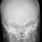
- mandible: in 75-80% of cases
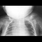
- clavicles
- scapula: 10% of cases
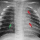
- ribs: lateral aspect; ipsilateral pleural effusion may appear
- calvaria
- ilia
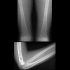
The ulnae are the long bones most commonly affected.
Lytic skull lesions have been reported.
The carpus, tarsus, phalanges and vertebral bodies are rarely involved.
Markers
Erythrocyte sedimentation rate (ESR), C-reactive protein (CRP), and alkaline phosphatase (ALP) levels are often elevated. The combination of the clinical picture, lab findings, and imaging findings is sufficient for a diagnosis.
Phases
Early, subacute, and late pathologic phases have been described, each with its accompanying radiologic features:
Early (acute)
- characterized by periostitis
- the inflammatory process may extend to adjacent soft tissues
- cortical resorption may occur
Subacute
- inflammation subsides
- periosteal thickening, with subsequent ossifying periostitis
- immature lamellar bone is deposited in layers under the periosteum, sometimes exuberantly
- osseous deposition may occur in adjacent soft tissues
Late
- removal of peripheral bone, from inner to outer surface
- may produce a thin-walled bone with a large medullary cavity in long-standing cases
- cortical remodeling may also occur
Long-term sequelae
- mandibular asymmetry and/or undergrowth
- persistent synostosis of the ribs: could cause scoliosis
- persistent synostosis of bones of the forearms and legs
- bowing of long bones
- leg length discrepancy
- disease recurrence in childhood
Radiographic features
Plain radiograph
May show all or some of the following :
- periosteal reaction, either single-layered or lamellated
- subperiosteal cortical hyperostosis
- dense laminated subperiosteal new bone formation
- marked increase in cortical width and density
- in the involved long bones, only the diaphysis is affected, sparing the metaphysis and epiphysis; consequently, the bone becomes spindle-shaped
- soft tissue swelling over the involved bones
- the mandible, clavicle, and ribs are most often affected, particularly in sporadic cases
Radiographic evidence of the disease may persist for years after resolution of clinical symptoms.
Ultrasound
Can identify soft tissue edema and early periosteal new bone formation. High-frequency transducers should be used.
MRI
Can show periostitis and soft tissue edema. It should be stressed that MRI usually does not offer much added-value in advancing the diagnosis unless infection or neoplasia are high on the differential list; indeed, at times, MRI appearance may confound the radiologist. Hence, radiography should be the primary modality of investigation and follow-up.
Nuclear imaging
During the active phase of the disease, there is markedly increased radiotracer uptake in the involved bones, both on bone and gallium (Ga-67) scans. The "bearded infant" appearance refers to intense radiotracer uptake in the mandible .
Nuclear scans can also be useful for showing the extent of skeletal involvement.
Treatment and prognosis
As noted above, Caffey disease is self-limiting and resolves spontaneously. Symptomatic treatment consists of NSAIDs, e.g. indomethacin.
History and etymology
Pediatric radiologist John Caffey (1895-1978) first described infantile cortical hyperostosis with colleague WA Silverman in 1945.
Differential diagnosis
- osteomyelitis: there are reports of association with Caffey disease
- non-accidental injury
- trauma
- physiologic periostitis: isolated to the tibiae, femora, and humeri
- skeletal dysplasias with osteosclerosis
- hypervitaminosis A
- hypovitaminosis C (scurvy)
- hyperphosphatemia
- prostaglandin E1 and E2 therapy (for intentionally maintaining ductus arteriosus patency)
- infection (e.g. syphilis, tuberculosis)
- Ewing sarcoma
- metastatic neuroblastoma
Other conditions can usually be excluded based on the narrow age range for the presentation of infantile cortical hyperostosis; the triad of irritability, swelling, and bone lesions; and the presence of mandibular involvement.
Siehe auch:
- Osteomyelitis
- Neuroblastom
- Ewing-Sarkom
- Battered-Child-Syndrom
- Hyperostose
- Knochen-in-Knochen-Aspekt
- Wiskott-Aldrich-Syndrom
- Kenny-Caffey-Syndrom
- vitamin A intoxication
- Hyperphosphatämie
und weiter:


 Assoziationen und Differentialdiagnosen zu Infantile kortikale Hyperostose: