calcific tendinitis


Calcific tendinitis (or calcific tendonitis) is a self-limiting condition due to the deposition of calcium hydroxyapatite within tendons, usually of the rotator cuff. It is a common presentation of the hydroxyapatite crystal deposition disease (HADD).
Epidemiology
Typically this condition affects middle-aged patients between the ages of 30 and 60, with a slight predilection for women .
Clinical presentation
The condition passes through four stages :
- asymptomatic
- fibrocartilaginous metaplasia (see below)
- symptoms are variable from none to pain on movement
- most symptomatic
- pain due to extravasation of calcium hydroxyapatite into adjacent tissues, especially subacromial bursa, causing calcific bursitis
- pain typically lasts two weeks
- variable symptomatology
- some restriction of movement common
- may last months
Pathology
Calcific tendinitis results from the deposition of calcium hydroxyapatite within the substance of a tendon and is thought to be due to decreased oxygen tension, leading to fibrocartilaginous metaplasia and secondary mineralization .
Location
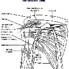
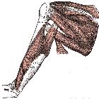
This condition most frequently affects the rotator cuff of the shoulder .
- supraspinatus: 80%
- infraspinatus: 15%
- subscapularis: 5%
- periarticular soft tissues in addition to tendons
- ligaments
- capsule
- bursae
However, the condition may occur anywhere in the body with the hip and knee being the other most common locations .
Radiographic features
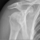
Plain radiograph
Calcific deposits are usually visualized as homogeneous hyperdensity with variable morphology, but typically globular/amorphous with smooth or ill-defined margins.
Ultrasound
Features of calcific tendinitis on ultrasound may include :
- a curvilinear/ovoid calcification with acoustic shadowing
- capsular soft tissue swelling
MRI
- T1
- hypointense homogeneous signal
- adjacent tendon may be thickened
- some enhancement surrounding deposit may be seen
- T2
- hypointense calcium deposits
- hyperintense signal may be present peripherally due to edema
- hyperintense subacromial-subdeltoid bursal fluid
- T2*: calcifications may bloom
Treatment and prognosis
Controversial and difficult to measure due to the inherent variability of the symptoms and the self-limiting nature of the disease. Potential treatments include :
- oral analgesic/anti-inflammatory medication
- subacromial local anesthetic/steroid injection
- aspiration of mineralized material
- ultrasound therapy
Differential diagnosis
In the shoulder consider:
- incidental calcification: seen in 2.5-20% of 'normal' healthy shoulders
- degenerative calcification
- seen in previously torn tendons
- generally smaller
- slightly older individuals
- loose bodies
- associated chondral defect
- associated secondary osteoarthritis
Siehe auch:
- periartikuläre Verkalkungen
- Hydroxylapatit-Ablagerungserkrankung
- Tendinosis calcarea der Rotatorenmanschette
- kalzifizierende Tendinitis des Musculus longus colli
- Bursa subacromialis
- Milwaukee-Schulter
- Tendinitis calcarea Musculus infraspinatus
- Rotatorenmanschette
- frozen shoulder
- Tendinose
- Tendinitis calcarea Musculus supraspinatus
- Tendinitis
- calcific tendinosis - pectoralis major
- Tendinosis calcarea Schulter
und weiter:


 Assoziationen und Differentialdiagnosen zu Tendinitis calcarea: