Hydroxylapatit-Ablagerungserkrankung


 nicht verwechseln mit: calcium pyrophosphate deposition disease (CPPD)
Hydroxyapatite crystal deposition disease (HADD) is a disease of uncertain etiology characterized by periarticular and intra-articular deposition of hydroxyapatite (HA) crystals.
The shoulder is the most frequently involved site with classic calcific tendinitis presentation.
Epidemiology
HADD is most commonly found in middle-aged individuals.
Clinical presentation
Localized pain is one of the primary manifestations of the disease, associated with swelling, tenderness, and variable limitation of joint motion. Often asymptomatic.
Pathology
The exact etiology has not been described; however, it is believed that HADD will begin to accumulate in damaged tendons (secondary to trauma) via fibrocartilaginous metaplasia .
The disease is characterized by calcium phosphate (calcium hydroxyapatite) crystal deposition in the periarticular soft tissues, especially in the tendons (best recognized as calcific tendinitis).
Most frequently it involves the shoulder joint, where crystal deposition occurs in the supraspinatus tendon, but the disease can affect numerous other sites as well. Calcific periarthritis or enthesitis would be the most appropriate term for this condition, as it occurs most commonly in the bony attachment of tendon near the joints .
HADD can affect other tendons of the body, such as the gluteus medius tendon, or along the femur, as well as at various sites of tendinous attachments (e.g. elbow, wrist, hand, knee, ankle, foot, and spine) .
Calcifications of varying sizes and shapes can involve the para-articular tendons, bursae, and capsule. The disease can be mono- or polyarticular.
Radiographic features
The specific appearance will vary based on the calcific stage, broken into the formative, resting and resorptive phases. The formative and resting phases will appear as round-to-ovoid calcification in the soft tissue with well-defined borders. The resorptive phase will appear ill-defined with a comet tail-like appearance. The resorptive may mimic a periosteal reaction.
Plain radiograph
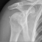
It appears as homogeneous, round-to-ovoid calcification in the soft tissue with well-defined or ill-defined margins. The most characteristic lesions are seen in the shoulder with supraspinatus and biceps tendon involvement, adjacent to the greater tubercle and the glenoid tubercle, respectively, where these tendons attach.
Deposits within the infraspinatus and teres minor tendons are not uncommon and can be seen adjacent to the greater tuberosity on internal rotation or axillary views .
Treatment and prognosis
Treatment is chiefly conservative, including NSAIDs, local heat application, and physiotherapy. Local corticosteroid injections may also be of benefit.
Surgical removal of calcifications may be appropriate for cases refractory to other attempts of conservative treatment.
Ultrasound ablation may prove to be of short-term benefit in particular cases. However, its long-term benefit has not yet been demonstrated .
Complications
When intra-articular, HA crystals can cause joint destruction. Any joint can be involved; the shoulder is most commonly affected, resulting in Milwaukee shoulder .
Differential diagnosis
The differential diagnosis of joint pain with calcification is extensive and occurs in many conditions such as:
- calcium pyrophosphate dihydrate deposition disease (CPPD)
- dystrophic calcification
- renal osteodystrophy
- hyperparathyroidism
- hypoparathyroidism
- tumoral calcinosis
- collagen vascular disease
- sarcoidosis
- ochronosis
- milk-alkali syndrome
- hypervitaminosis D
Another consideration for a calcific opacity in proximity to a joint (typically knee, elbow, etc.) would be a loose body from an osteochondritis dissecans (OCD) donor site. Therefore, radiographic findings, a thorough history, as well as physical examination, appropriate lab tests and additional imaging modalities, should be utilized to identify the most likely etiology .
Siehe auch:
- Weichteilverkalkungen
- Tendinitis calcarea
- Tendinosis calcarea der Rotatorenmanschette
- kalzifizierende Tendinitis des Musculus longus colli
- Bursa subacromialis
- Milwaukee-Schulter
- Tendinitis calcarea Musculus infraspinatus
- Kalziumpyrophosphat-Ablagerungskrankheit
- frozen shoulder
- Tendinose
- Bursitis calcarea
- Tendinitis calcarea Musculus supraspinatus
- Kristallarthropathie
- calcific tendinosis - pectoralis major
- calcific periarthritis
- Tendinosis calcarea Schulter
und weiter:


 Assoziationen und Differentialdiagnosen zu Hydroxylapatit-Ablagerungserkrankung:


