calyceal diverticulum


Calyceal diverticula, also known as pyelocalyceal diverticula are congenital outpouchings from the renal calyx or pelvis into the renal cortex. These diverticula are lined with transitional cell epithelium.
Epidemiology
Relatively uncommon, seen in 0.21% to 0.60% of intravenous urograms (IVU).
Associations
- gender: females are more commonly affected than men, with a ratio of 2:1
- high incidence of stone formation due to urine stagnation.
Clinical presentation
The majority of cases are asymptomatic and discovered incidentally on imaging. Up to 50%, though, present with hematuria, calculi, flank pain and/or recurring infection.
Pathology
The most common theory for the origin of calyceal diverticula is a failure of regression of the third and fourth-generation ureteric buds, as a result of obstructing stones or infection.
There are two categories of calyceal diverticula:
- type I: more common, communicates with a minor calyx
- type II: communicates with a major calyx or the renal pelvis and tends to be symptomatic
Radiographic features
Plain radiograph
Only a diverticulum containing milk of calcium will be visible on a plain radiograph. It appears as a meniscus-shaped density on an upright radiograph that changes its shape with changing position, i.e. either supine or decubitus radiographs.
Ultrasound
Calculi or layered dependent milk of calcium complicating a calyceal diverticulum appear echogenic on ultrasound, but only the former will cast an acoustic shadow.
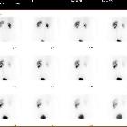
CT
On nephrographic phase contrast-enhanced CT, a calyceal diverticulum will have an appearance similar to that of a simple cyst. The diagnosis is made with certainty in the excretory phase when the cystic structure fills with contrast material due to communication with the collecting system, and layering of contrast material is seen within. This helps differentiate it from a renal cyst, which does not connect with the collecting system.
A calyceal diverticulum complicated by calculi or layered dependent milk of calcium will be hyperattenuating on an unenhanced CT.
Differential diagnosis
General imaging differential considerations include
- renal milk of calcium cysts
- simple renal cyst
Siehe auch:
und weiter:


 Assoziationen und Differentialdiagnosen zu Kelchdivertikel: