cerebral atrophy


Cerebral atrophy is the morphological presentation of brain parenchymal volume loss that is frequently seen on cross-sectional imaging. Rather than being a primary diagnosis, it is the common endpoint for range disease processes that affect the central nervous system. Though often no identifiable cause is found, certain patterns of atrophy can be helpful in certain clinical scenarios, most notably in neurodegenerative diseases.
It is a common finding in the elderly population, and so there is some controversy as to when imaging changes are labeled as cerebral atrophy, rather than simply "involutional" or "age-related" when the patient has normal cognition.
Terminology
As cerebral atrophy is simply the compensatory enlargement of the CSF spaces from reducing brain parenchymal volume, it is akin to hydrocephalus ex vacuo. However, the later term is usually reserved when referring to focal volume loss in the brain following a pathological insult (i.e. hemorrhage) rather than the often idiopathic more generalized changes seen with age.
Clinical presentation
As it is not a distinct disease entity, there is no uniform mode of presentation and the finding of atrophy is often incidental when imaging is taken for some other indication.
Cognitive dysfunction and acute confusion are common reasons patients with atrophy may undergo imaging.
Pathology
The underlying pathological causes can be broadly distinguished based on whether the atrophy is focal or generalized:
- generalized atrophy
- age-related
- cerebrovascular disease
- end-stage multiple sclerosis
- alcohol abuse (cerebellar vermian atrophy)
- drug abuse
- post-traumatic (diffuse axonal injury)
- post-infective (eg. meningitis)
- certain neurodegenerative diseases
- progressive supranuclear palsy
- focal atrophy
- post-ischemic injury
- post-traumatic (hemorrhage, contusion)
- certain neurodegenerative disease
Radiographic features
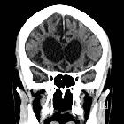
CT and MRI are equally able to demonstrate cortical atrophy, but MRI is more sensitive in detecting focal atrophic changes in the nuclei. Characteristic features include prominent cerebral sulci (i.e. cortical atrophy) and ventriculomegaly (i.e. central atrophy) without bulging of the third ventricular recesses. It can be difficult to distinguish this from the changes seen in normal pressure hydrocephalus.
Certain important patterns of cerebral atrophy that are more specific include:
- severe frontal and anterior temporal
- Pick disease
- head of caudate nuclei
- Huntington disease
- posterior parietal and frontal
- corticobasal degeneration
- atrophy of tectum, globus pallidus, and frontal lobes
- progressive supranuclear palsy
- generalized with atrophy of substantia nigra
- Parkinson disease
- severe hippocampal atrophy
- Alzheimer dementia
Differential diagnosis
Siehe auch:
- Kleinhirnatrophie
- einseitige Hirnatrophie
- frontale Atrophie
- zerebrale Atrophie quantifizieren
- parietale Hirnatrophie
- posterior cerebral atrophy
und weiter:


 Assoziationen und Differentialdiagnosen zu Hirnatrophie:
