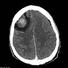
chorioadenoma destruens
Invasive mole is a tumorous growth associated with gestation and falls under the spectrum of gestational trophoblastic disease. Due to their aggressive growth characteristics, invasive moles are considered locally invasive non-metastasizing neoplasms.
Epidemiology
An invasive mole develops in approximately 10-20% of patients after molar evacuation and infrequently after other gestations. It is defined as a mole that penetrates and may even perforate the uterine wall.
Pathology
Invasive moles arise from hydatidiform moles. There is an invasion of the myometrium by hydropic chorionic villi, accompanied by the proliferation of trophoblast. A tumor is locally destructive and may invade parametrial tissue and blood vessels.
Markers
As with other forms of the gestational trophoblastic disease, maternal serum βHCG values are markedly elevated.
Radiographic features
Often it can be difficult to differentiate an invasive mole from other forms of gestational trophoblastic disease in imaging.
Pelvic ultrasound
May be seen as an echogenic vascular mass invading the myometrium. Color Doppler interrogation will show high velocity, low impedance flow.
Pelvic MRI
On MRI, it often appears as a poorly defined mass that deeply invades the myometrium. Complete or partial disruption of the junctional zone may also be seen.
Typical signal characteristics include:
- T1: isointense to the myometrium with scattered foci of high signal intensity (from the presence of hemorrhage)
- T2: mixed signal intensity
Molar-like structures appear as tiny cystic lesions within the well-enhanced zone of trophoblastic proliferation in a mass of the invasive mole.
With the penetration of a tumor into the myometrium, the invasive mole can appear as a more aggressive entity compared with a choriocarcinoma.
Siehe auch:
und weiter:

 Assoziationen und Differentialdiagnosen zu invasive mole:

