cystic bronchiectasis

Cystic
bronchiectasis • Morphological types of bronchiectasis (illustration) - Ganzer Fall bei Radiopaedia


Cystic
bronchiectasis • Cystic bronchiectasis - Ganzer Fall bei Radiopaedia

Cystic
bronchiectasis • Cystic bronchiectasis - Ganzer Fall bei Radiopaedia

Cystic
bronchiectasis • Cystic bronchiectasis - Ganzer Fall bei Radiopaedia

Cystic
bronchiectasis • Cystic bronchiectasis - Ganzer Fall bei Radiopaedia

Cystic
bronchiectasis • Cystic bronchiectasis - Ganzer Fall bei Radiopaedia

Cystic
bronchiectasis • Cystic bronchiectasis - Ganzer Fall bei Radiopaedia

Cystic
bronchiectasis • Cystic bronchiectasis - Ganzer Fall bei Radiopaedia

Cystic
bronchiectasis • Cystic bronchiectasis - Ganzer Fall bei Radiopaedia

Cystic
bronchiectasis • Cystic bronchiectasis - Ganzer Fall bei Radiopaedia


Cystic
bronchiectasis • Cystic bronchiectasis - Ganzer Fall bei Radiopaedia

Cystic
bronchiectasis • Cystic bronchiectasis - Ganzer Fall bei Radiopaedia

Cystic
bronchiectasis • Cystic bronchiectasis - cystic fibrosis - Ganzer Fall bei Radiopaedia

Cystic
bronchiectasis • Cystic bronchiectasis - Ganzer Fall bei Radiopaedia

Beyond
bronchitis: a review of the congenital and acquired abnormalities of the bronchus. Williams-Campbell syndrome. a Frontal chest x-ray demonstrating bilateral architectural distortion and coarse interstitial markings with cystic appearing regions in the lower lobe consistent with the scarring and diffuse saccular bronchiectasis seen in Williams-Campbell syndrome. b Coronal CT imaging demonstrating the severe bilateral cystic bronchiectasis of the subsegmental (4th-6th generation) bronchi commonly seen in Williams-Campbell syndrome

Kartageners
syndrome. Coronal and axial images of trilobed left lung and bilobed right lung. Yellow arrow shows horizontal fissure in left lung

Kartageners
syndrome. Cystic bronchiectasis in both lungs with predominance in lower lobes. Few centrilobular micro nodules representing mucoid impaction with few areas of ground glass opacities are seen in the left lower lobe.

Cystic
bronchiectasis • Cystic bronchiectasis - brunch of grapes appearance - Ganzer Fall bei Radiopaedia
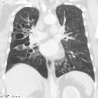
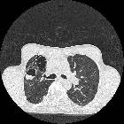
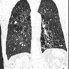
Cystic bronchiectasis is one of the less common morphological forms of bronchiectasis. It may be present on its own or may occur in combination with other forms of bronchiectasis.
For a general discussion, please refer to the article on bronchiectasis.
Radiographic features
It is characterized by saccular dilatation of bronchi that extends to the pleural surfaces. When aggregated these may give a "bunch of grapes" like appearance.
Siehe auch:
- Bronchiektasen
- multiple zystische Lungenherde
- Kartagener-Syndrom
- primäre ciliäre Dyskinesie
- congenital cystic bronchiectasis
und weiter:

 Assoziationen und Differentialdiagnosen zu cystic bronchiectasis:


