Dextrokardie


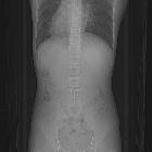
Dextrocardia is a congenital cardiac malrotation in which the heart is situated on the right side of the body (dextroversion) with the apex pointing to the right.
Terminology
Dextrocardia merely refers to the laterality of the heart, it says nothing about the orientation of the patient's other organs. Laevocardia is the term used for the heart being positioned on the left as is found in the vast majority of the healthy population.
Epidemiology
Dextrocardia is believed to occur in approximately 1 in 12,000 people .
Pathology
There are two main types of dextrocardia:
- dextrocardia of embryonic arrest (also known as isolated dextrocardia)
- the heart is simply placed further right in the thorax than is normal
- commonly associated with severe defects of the heart, including abnormalities such as pulmonary hypoplasia
- dextrocardia with situs inversus
- dextrocardia situs inversus refers to the heart being a mirror image situated on the right side
- for all visceral organs to be mirrored, the correct term is dextrocardia situs inversus totalis
- although persons with dextrocardia situs inversus tend not have any medical problems from the disorder, some are prone to a number of bowel, esophageal, bronchial and cardiac problems where some of these conditions can be life-threatening if uncorrected
Associations
Diagnosis
ECG
The most prominent abnormality recognized is often the "global inversion" of standard (limb) lead I, with inversion of the P wave, QRS complex, and T wave, suggestive of either dextrocardia or limb lead misplacement (the RA, or right arm, lead was placed on the left and vice versa). Use of the precordial leads allows discrimination of these two entities, specifically by observing the precordial R wave progression.
As one progresses from the right (represented by leads V1-V3) through the left precordium (leads V4-6), a progressive increase in the amplitude of the (first positive QRS deflection) R wave is expected, with poor R wave progression (PRWP) diagnosed when the amplitude of the R wave in V3 is <3 mm (often suggestive of an anteroseptal myocardial infarction of indeterminate age).
Misplaced limb leads will not affect the precordium, therefore a normal R wave progression is expected. Dextrocardia, however, will demonstrate either loss of normal R wave progression (and consequently, PRWP) or a reversal of the normal progression, with dominant S waves present throughout the precordium and R waves decreasing in positivity progressively from right to left. Other common ECG features include:
- right axis deviation (normal axis between -30 and +90 degrees)
- QRS positivity in lead aVR (usually predominantly negative)
- reversal of pattern with inversion of limb leads and precordial leads
Differential diagnosis
- spurious dextrocardia (see below)
Practical points
When approaching the chest radiograph in someone that appears to have dextrocardia it is important to exclude spurious dextrocardia first. This is by far the commonest cause of a dextrocardia-like appearance and is a technical error due to the radiographer/technician inadvertently flipping the image over in the mediolateral plane when processing the image. Therefore it is important to check the orientation marker on the image; usually in a spurious dextrocardia the marker will be on the 'wrong' side i.e. left marker on the right, or vice-versa.
ECG leads must be placed in reversed positions on a person with dextrocardia. In addition, when defibrillating someone with dextrocardia, the pads should be placed in reverse positions. That is, instead of upper right and lower left, pads should be placed upper left and lower right.
Siehe auch:
und weiter:

 Assoziationen und Differentialdiagnosen zu Dextrokardie:


