Dieulafoy lesion


Dieulafoy lesions (also known as exulceratio simplex) are uncommon but important causes of acute upper gastrointestinal bleeding. A Dieulafoy lesion is characterized by a dilated tortuous submucosal artery that erodes overlying gastrointestinal mucosa most commonly found in the stomach.
Epidemiology
- contributes to ~1.5% of all acute gastrointestinal bleeding
- male : female 2:1
- can occur at any age but presents more commonly in older patients
Clinical presentation
Patients present with hematemesis, which can be massive, and/or melena .
Pathology
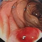
Dieulafoy lesions are dilated and tortuous submucosal arteries that erode the overlying gastrointestinal mucosa and result in bleeding .
Location
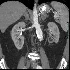
Although they can be present throughout the gastrointestinal tract, approximately 70% are located in the stomach.
- Gastric: In the stomach, the lesser curvature is the commonest location
- Extragastric: in descending order of frequency, appearance in the duodenum, right colon, esophagus, rectum, and anal canal have been described
- rare cases outside the GI tract entirely, e.g. bronchus
Radiographic features
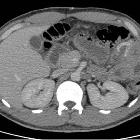
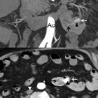
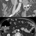
CT
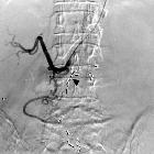
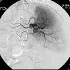
CT angiogram may show:
- enlarged submucosal vessel with or without active contrast extravasation
- vessels can appear linear or serpentine or as a non-specific blush of mucosal/submucosal contrast
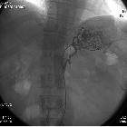
Treatment and prognosis
Endoscopic treatment is the treatment of choice and has reported success rate over 90% . Angiography plus embolization or surgery can be considered in refractory cases.
History and etymology
It was first described by M T Gallard a French surgeon in 1884 but was described in more detail by Paul Georges Dieulafoy (1839-1911) , another French surgeon in 1898 .
Siehe auch:


 Assoziationen und Differentialdiagnosen zu Dieulafoy-Ulcus: