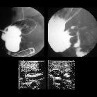
Duodenalstenose

Ösophagusatresie
Typ III B und Duodenalstenose bei Pancreas anulare mit typischem "Double bubble sign". Rechts mehr als links infiltratverdächtige Verdichtungen.

Palliative
Metallstents im DHC und Duodenum bei Stenose durch Pankreaskarzinom

Duodenal
lymphoma: A rare cause of obstructive jaundice. Homogeneous hypodense circumferential thickening of the duodenal wall (arrows), with loco-regional adenopathies (arrowheads).

Peptic
duodenal stricture causing gastric outlet obstruction: CT diagnosis with endoscopic correlation. Upright radiograph showed overdistended stomach (*) with intraluminal stagnant fluid despite recent fasting (note air-fluid levels indicated by thick arrows).

Peptic
duodenal stricture causing gastric outlet obstruction: CT diagnosis with endoscopic correlation. Coronal (c, detail d) and axial (detail e) images confirmed overdistended stomach with stagnant fluid (*). The pylorus and proximal duodenum showed circumferential hypoenhancing mural thickening (+) and depressed focal thinning of enhancing mucosal contour (thin arrows).

Peptic
duodenal stricture causing gastric outlet obstruction: CT diagnosis with endoscopic correlation. Coronal images (c, detail d) confirmed overdistended stomach with stagnant fluid (*). The pylorus and proximal duodenum showed circumferential hypoenhancing mural thickening (+) and depressed focal thinning of enhancing mucosal contour (thin arrows).

Peptic
duodenal stricture causing gastric outlet obstruction: CT diagnosis with endoscopic correlation. Coronal (c, detail d) and axial (detail e) images confirmed overdistended stomach with stagnant fluid (*). The pylorus and proximal duodenum showed circumferential hypoenhancing mural thickening (+) and depressed focal thinning of enhancing mucosal contour (thin arrows).

Peptic
duodenal stricture causing gastric outlet obstruction: CT diagnosis with endoscopic correlation. Sagittal images (f, detail g) images confirmed overdistended stomach with stagnant fluid (*). The contracted pylorus and proximal duodenum (arrowheads) showed hypoenhancing mural thickening (+), depressed focal thinning of enhancing mucosal contour (thin arrows). Superior duodenal flexure (§).

Peptic
duodenal stricture causing gastric outlet obstruction: CT diagnosis with endoscopic correlation. Sagittal images (f, detail g) images confirmed overdistended stomach with stagnant fluid (*). The contracted pylorus and proximal duodenum (arrowheads) showed hypoenhancing mural thickening (+) and depressed focal thinning of enhancing mucosal contour (thin arrows).

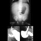
Newborn with
vomiting. AXR AP obtained after gastric decompression via nasogastric tube (above) shows gas present through the bowel. AP image from an upper GI exam (below) shows the stomach and first and second parts of the duodenum to be extremely dilated. On a delayed image obtained 15 minutes later, a small amount of contrast was seen to have passed out of the duodenum into the small bowel.The diagnosis was duodenal stenosis.
 Assoziationen und Differentialdiagnosen zu Duodenalstenose:
Arteria-mesenterica-superior-Syndrom


