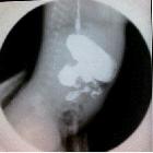
Stenose Jejunum

Newborn with
vomiting who had a triple bubble sign on prenatal US. Early (above) image from an UGI exam shows dilation of the stomach, duodenum, and proximal jejunum. Later image from the same exam (below) shows contrast progressing through the remainder of the jejunum, which is of a normal caliber.The diagnosis was jejunal stenosis.

Ultrasonographic
diagnosis of congenital membranous jejunal stenosis and gastric duplication cyst in a newborn: a case report. Upper gastrointestinal contrast study showing marked dilatation of the duodenum

Ultrasonographic
diagnosis of congenital membranous jejunal stenosis and gastric duplication cyst in a newborn: a case report. Ultrasonography showing dilatation of the duodenum and proximal jejunum. A membrane (arrow) and a pinhole (small arrowhead) are seen in the jejunal membrane
Stenose Jejunum
Siehe auch:

 Assoziationen und Differentialdiagnosen zu Stenose Jejunum:


