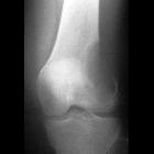
einfache (juvenile) Knochenzyste Femur


einfache (juvenile) Knochenzyste Femur
einfache (juvenile) Knochenzyste Radiopaedia • CC-by-nc-sa 3.0 • de
Unicameral bone cysts (UBC), also known as simple bone cysts, are common benign non-neoplastic lucent bony lesions that are seen mainly in childhood and typically remain asymptomatic. They account for the S (simple bone cyst) in FEGNOMASHIC, the commonly used mnemonic for lytic bone lesions.
Epidemiology
They are usually found in children in the 1st and 2nd decades (65% in teenagers) and are more common in males (M:F ~ 2-3:1) .
Clinical presentation
These lesions are usually asymptomatic and found incidentally, although pain, swelling and stiffness of the adjacent joint also occur. The most frequent complication is a pathological fracture, and this is frequently the cause of presentation .
Pathology
When uncomplicated by fracture the cysts contain clear serosanguineous fluid surrounded by a fibrous membranous lining. It is thought to arise as a defect during bone growth which fills with fluid, resulting in expansion and thinning of the overlying bone.
During the active phase, the cyst remains adjacent to the growth plate. As the lesion becomes inactive it migrates away from the growth plate (normal bone is formed between it and the growth plate) and it gradually resolves .
Location
They are typically intramedullary and are most frequently found in the metaphysis of long bones, abutting the growth plate . Locations include :
- proximal humerus: most common 50-60%
- proximal femur: 30%
- other long bones
- occurrence elsewhere is relatively uncommon, and usually occurs in adults
- spine: usually posterior elements
- pelvis: only 2% of UBC
UBCs can be rarely seen in adults in unusual locations such as in the talus, calcaneus, or the iliac wing.
Radiographic features
Plain radiograph
UBCs are well defined geographic lucent lesions with a narrow zone of transition, mostly seen in skeletally immature patients, which are centrally located and show a sclerotic margin in the majority of cases with no periosteal reaction or soft tissue component. They sometimes expand the bone with thinning of the endosteum without any breach of the cortex unless there is a pathologic fracture. Prominent ridges of bone can appear as pseudotrabeculation on x-ray but in fact, UBC is made of one contiguous cystic space. Rarely, they are truly multiloculated .
If there is fracture through this lesion a dependent bony fragment may be seen, and this is known as the fallen fragment sign.
CT and MRI
CT and MRI add little to the diagnosis, but are however helpful in eliminating other entities that can potentially mimic a simple bone cyst (see differential diagnosis below).
MR signal characteristics for an uncomplicated lesion include:
- T1: low signal
- T2: high signal
Usually there no fluid-fluid levels unless there has been a complication with hemorrhage.
Scintigraphy
Unicameral bone cyst on bone scintigraphy tends to appear as foci of photopenia (cold spot). However, a pathological fracture would cause an increased radioisotope activity.
Treatment and prognosis
Intervention is usually not required for an asymptomatic lesion. If large and threatening to fracture, or causing deformity then an intralesional steroid injection can be performed . If fractured the bone usually heals normally . In some instances, surgery with curettage and bone grafting is required.
Differential diagnosis
General imaging differential considerations include
- intraosseous lipoma
- fibrous dysplasia
- eosinophilic granuloma (EG)
- giant cell tumor of bone: usually older, extending to articular surface
- non ossifying fibroma: eccentric, cortical base
- haemophilic pseudotumor (intraosseous)
- aneurysmal bone cyst (ABC): usually eccentric
See also
Siehe auch:

 Assoziationen und Differentialdiagnosen zu einfache (juvenile) Knochenzyste Femur:


