eingeblutete Leberzyste

Cystic liver
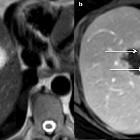
lesions: a pictorial review. Bilobulated haemorrhagic hepatic cyst in a 73-year-old female. a Coronal non-enhanced computed tomography shows a bilobulated cyst with focal hyperattenuation in the inferior part (arrow); b coronal T2-weighted magnetic resonance imaging shows heterogeneous hypointensity inside the inferior part of the cyst (arrow). c Contrast-enhanced ultrasonography confirms the non-enhancement of the cystic part of the lesion (arrowhead)

Cystic liver
lesions: a pictorial review. Subacute haemorrhage in a simple hepatic cyst in a 70-year-old male. Ultrasonography shows a spontaneous mobile area of hyperechogenicity inside the cyst, appearing as a “fern leaf”

Cystic liver
lesions: a pictorial review. Haemorrhagic cyst in a 55-year-old male patient. a Axial T1 fat-sat-weighted magnetic resonance imaging shows hyperintense lesion and (b) axial T2-weighted magnetic resonance imaging shows a heterogeneous hyperintensity; c and d axial T1-fat-sat-weighted imaging without and with subtraction shows no enhancement after gadolinium-chelate injection


 Assoziationen und Differentialdiagnosen zu eingeblutete Leberzyste: