Differenzialdiagnosen zystischer Leberläsionen

Cystic liver
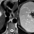
lesions: a pictorial review. Typical non-invasive mucinous cystic neoplasm of the liver in a 59-year-old female. a Axial T2-weighted magnetic resonance imaging shows a solitary hyperintense cystic lesion, with hypointense septa (arrows). b Axial T1-weighted magnetic resonance imaging on portal venous phase depicts the enhancement of the capsule and septa (arrows)

Cystic liver
lesions: a pictorial review. Bile duct hamartomas in a 52-year-old female. a Multiple hyperechoic round nodules (arrows) at ultrasonography, displaying a “snowstorm” pattern. b Coronal magnetic resonance cholangiopancreatography shows several small hyperintense nodules, which do not communicate with the biliary tract (arrows). c Axial computed tomography on portal venous phase shows bilobar small round-shaped hypoattenuating nodules, which can be better detected with an axial magnetic resonance cholangiopancreatography sequence (d)

Cystic liver
lesions: a pictorial review. Caroli syndrome in a 7-year-old female, at ultrasonography. Several round anechoic cysts (stars) with thin wall, increased through-transmission, connected to the dilated fusiform biliary tract (arrows), some of which show the highly specific “central dot sign” (arrowhead)

Cystic liver
lesions: a pictorial review. Very large simple hepatic cyst in a symptomatic 64-year-old female with abdominal pain. a Axial and (b) coronal computed tomography on portal venous phase shows a very large simple hepatic cyst. The cyst had grown progressively for a certain number of years and was eventually treated by laparoscopic fenestration, resulting in pain relief

Cystic liver
lesions: a pictorial review. Bilobulated haemorrhagic hepatic cyst in a 73-year-old female. a Coronal non-enhanced computed tomography shows a bilobulated cyst with focal hyperattenuation in the inferior part (arrow); b coronal T2-weighted magnetic resonance imaging shows heterogeneous hypointensity inside the inferior part of the cyst (arrow). c Contrast-enhanced ultrasonography confirms the non-enhancement of the cystic part of the lesion (arrowhead)

Cystic liver
lesions: a pictorial review. Infected simple hepatic cysts in a 77-year-old male with autosomal dominant polycystic liver disease. a Axial non-enhanced computed tomography shows two cysts under pressure (arrow), slightly more attenuating than the non-infected cysts (arrowhead); b with a thick enhanced wall on the portal venous phase (arrowhead)

Cystic liver
lesions: a pictorial review. Haemorrhagic cyst in a 55-year-old male patient. a Axial T1 fat-sat-weighted magnetic resonance imaging shows hyperintense lesion and (b) axial T2-weighted magnetic resonance imaging shows a heterogeneous hyperintensity; c and d axial T1-fat-sat-weighted imaging without and with subtraction shows no enhancement after gadolinium-chelate injection

Cystic liver
lesions: a pictorial review. Rupture of a hepatic cyst in a 89-year-old male with severe abdominal pain. Axial computed tomography on portal venous phase demonstrates a floating wall (arrow) associated with pericystic and peri-hepatic fluid collection

Cystic liver
lesions: a pictorial review. Caroli disease in a 24-year-old male. a Axial magnetic resonance imaging in T2 and (b) in T1 on portal venous phase show both fusiform (arrows) and connected round (arrowheads) dilatations, better depicted on magnetic resonance cholangiopancreatography images (c). There is no associated sign of fibrosis

Cystic liver
lesions: a pictorial review. Caroli syndrome in a 53-year-old female. a Axial magnetic resonance imaging on portal venous phase shows peripheric fusiform dilated bile duct, hyperintense on axial T2-weighted magnetic resonance imaging (b), with the specific “central dot sign” (arrows). Note the presence of spleno-renal shunts due to portal hypertension

Cystic liver
lesions: a pictorial review. Schematic figure (axial section of a liver) showing the different types of ductal plate malformation. The size of the affected ducts is closely correlated to the ductal development phase

Cystic liver
lesions: a pictorial review. Hepatic alveolar echinococcosis in a 40-year-old male. a Ultrasonography shows multiple round hyperechoic nodules (arrows) with cystic anechoic parts (arrowhead). b On Axial T2-weighted magnetic resonance imaging, the lesions appear as multivesicular cystic masses (arrows) with a fibrous component (arrowhead). c and d On axial non-enhanced T1-fat-sat-weighted magnetic resonance imaging, the fibrous part is enhancing on late phase (arrow)

Cystic
lesions of the liver (differential) • Cystic hepatic metastasis - Ganzer Fall bei Radiopaedia

Cystic
lesions of the liver (differential) • Simple hepatic cyst - Ganzer Fall bei Radiopaedia

Cystic
lesions of the liver (differential) • Von Meyenburg complex - Ganzer Fall bei Radiopaedia

Cystic
lesions of the liver (differential) • Polycystic liver - Ganzer Fall bei Radiopaedia

Cystic
lesions of the liver (differential) • Hydatid cyst of the liver - Ganzer Fall bei Radiopaedia

Cystic
lesions of the liver (differential) • Pyogenic liver abscess - Ganzer Fall bei Radiopaedia

Cystic
lesions of the liver (differential) • Biliary cystadenoma - Ganzer Fall bei Radiopaedia

Cystic liver
lesions: a pictorial review. Incidentally found simple hepatic cyst in an asymptomatic 64-year-old male. a Ultrasonography shows a round homogeneous anechoic cystic hepatic lesion, well-circumscribed and without any mural nodule or vegetation. The arrows show an increased through transmission, confirming the cystic nature of the lesion. b T2-weighted magnetic resonance imaging shows a highly hyperintense round lesion. c and d Computed tomography without and with contrast on portal venous phase shows a round homogeneous non-enhancing hypoattenuating lesion (3 Hounsfield Units)

Cystic liver
lesions: a pictorial review. Simple algorithms helping to delineate diagnosis according to the number of cysts

Leberzysten
in der Computertomographie: Gleiche Dichte wie Flüssigkeit (siehe Aszitessaum neben der Leber).

Cystic liver
lesions: a pictorial review. Cystic metastasis from malignant melanoma in a 72-year-old female. a, b Axial computed tomography on arterial and portal venous phases shows unique hypoattenuating (20 Hounsfield Units) nodule of the right liver lobe, too small to be characterised. c Contrast-enhanced ultrasonography displays an enhanced wall as a “rim” (arrows)

Cystic liver
lesions: a pictorial review. Autosomal dominant polycystic kidney disease in a 38-year-old male patient. a Axial computed tomography on portal venous phase and b axial T2-weighted magnetic resonance imaging show several simple hepatic cysts and also numerous kidney cysts (arrows), with fluid signal (hypodense/hyperintense on T2)

Cystic liver
lesions: a pictorial review. cyst echinococcosis 2, in a 50-year-old male. a Axial non-enhanced computed tomography shows a heterogeneous mass with a cystic part (arrow) and calcification of the wall (arrowhead). b On axial computed tomography on portal venous phase no enhancement of the cystic component is observed. c The cystic part and the differentiation between the daughter (arrow) and the mother (star) cysts are better evaluated on axial T2-weighted magnetic resonance imaging. d Coronal 3D magnetic resonance cholangiopancreatography excludes biliary fistula from the possible diagnosis

Cystic liver
lesions: a pictorial review. Complicated hydatid cyst in a 50-year-old female. Imaging displays a cyst very close to the biliary tract with lobulated margins, showing a loss of intralesional pressure, most probably resulting from a complication of the biliary connection (not found on magnetic resonance cholangiopancreatography). a Axial T2-weighted magnetic resonance imaging. b Coronal 3D magnetic resonance cholangiopancreatography. c and d Axial T1-fat-sat-weighted imaging without and with contrast on portal venous phase

Cystic liver
lesions: a pictorial review. Hepatic pyogenic abscesses in a 49-year-old male. a Ultrasonography shows a large heterogeneous hypoechoic lesion, with anechoic parts (star) and increased through transmission (arrows) confirming its cystic nature. b and c Axial computed tomography on portal venous phase displays multiple abscesses appearing as cystic masses, with a specific sign: the double target (arrows). Note the bubble of gas in one of them (arrow-head)

Cystic liver
lesions: a pictorial review. Escherichia Coli abscess in an 89-year-old male. Axial computed tomography on portal venous phase shows three small hypoattenuating areas (arrows) forming the “cluster sign” and a large central hypoattenuating cavity (star)

Cystic liver
lesions: a pictorial review. Cystic metastasis of a mucinous tumour of the rectum in a 72-year-old male. The hyperintensity on axial T2-weighted magnetic resonance imaging a is marked but the multiple tiny septa (arrows) incompletely enhanced on portal venous phase (b) are sufficient to reach a conclusive diagnosis in this the context

Cystic liver
lesions: a pictorial review. Pure cystic metastasis of a neuroendocrine tumour in a 69-year-old female. The hyperintensity on axial T2-weighted magnetic resonance imaging (a) is marked and there is no enhancement on arterial (arrow) or portal venous (arrow-head) phase (b, c)

Cystic liver
lesions: a pictorial review. Metastatic pancreatic neuroendocrine tumour in a 60-year-old male. a Axial computed tomography on portal venous phase shows a lobulated hypodense cystic nodule in segment V with enhanced mural nodule and septa (arrow), very hyperintense on T2-weighted magnetic resonance imaging (b). A second cystic subcapsular nodule is visible in segment VI

Cystic liver
lesions: a pictorial review. Intraductal papillary neoplasm of the bile duct in a 67-year-old female. a Axial T2-weighted magnetic resonance imaging displays intraductal heterogeneous cystic and tissular mass (star) with dilated bile duct in contact (arrows). The cystic part of the mass is better seen at magnetic resonance cholangiopancreatography (b), and so are the upstream biliary dilatation (arrows) and the connection to the biliary tree (arrowhead). c and d Axial T1-weighted non-enhanced magnetic resonance imaging on arterial phase shows a highly enhanced tissular part

Zystenleber
mit zum Teil etwas dichteangehobenen, mutmaßlich proteinreicheren Zysten. Ein Teil der Zysten auch randständig etwas verkalkt. Bei dem Patienten fanden sich allenfalls minimale, kleine Zysten in den Nieren und anderen Organen, so dass sich nicht das Bild einer autosomal-dominanten polyzystische Nierenerkrankung (ADPKD) ergab, sondern eher das einer ("isolierten") polyzystischen Lebererkrankung (PCLD). Eine genetische Abklärung konnte jedoch nicht erfolgen.

Cystic liver
lesions: a pictorial review. Invasive mucinous cystic neoplasm of the liver in a 50-year-old female. a Axial non-enhanced computed tomography shows a hypoattenuating lesion with a thick heterogeneous enhancing wall on portal venous phase (b), better seen on T1-weighted magnetic resonance imaging on portal venous phase (d) (arrows). Note the presence of cystic portions on axial T2-weighted sequence (c), in very intense hypersignal T2 (arrows)

Cystic liver
lesions: a pictorial review. Ciliated hepatic foregut duplication cyst in a 15-year-old male. a Axial non-enhanced computed tomography shows a subcapsular nodule of the fourth segment with spontaneous attenuation value of around 50 Hounsfield Units. b On axial T1-weighted magnetic resonance imaging, it is spontaneously hyperintense. c It displays a high level of hyperintensity on axial T2-weighted magnetic resonance imaging. d Axial T1-weighted magnetic resonance imaging on portal venous phase shows neither contrast enhancement nor wall thickening

Cystic liver
lesions: a pictorial review. Spontaneous disappearance of a cystic lesion in a 5-year-old boy between 2017 and 2018, which led to the diagnosis of this lesion as a most likely liver lymphatic malformation. a Ultrasonography shows biloculated homogeneous cyst (arrow), which vanished one year later (star). It displayed fluid signal (arrows) on axial T2-weighted (b) and T1-weighted magnetic resonance imaging in 2017 (c). The main differential diagnosis of this lesion would be two fused simple hepatic cysts or mesenchymal hamartoma

Cystic liver
lesions: a pictorial review. Peribiliary cyst in a 69-year-old cirrhotic male. a Ultrasonography shows multiple periportal (arrowhead) cysts (arrows). b The portal vessels are clearly visible (arrowhead) on axial portal venous phase magnetic resonance imaging, showing the periportal location of the cysts. c and d Axial T2-weighted magnetic resonance imaging and magnetic resonance cholangiopancreatography images show multiple small cysts along thin, non-dilated bile ducts

Cystic liver
lesions: a pictorial review. Infected cyst in a 65-year-old male patient with autosomal dominant polycystic liver disease. a Axial non-enhanced computed tomography depicts one more attenuating cyst among multiple simple cysts (cross); b 18-F-FDG PET imaging shows the increased metabolism of that cyst (SUV at 5,1)

Leberabszess
in der Computertomographie axial: Man erkennt die hypodense Läsion in der Leber mit einem Randwall.

Cystic liver
lesions: a pictorial review. Subacute haemorrhage in a simple hepatic cyst in a 70-year-old male. Ultrasonography shows a spontaneous mobile area of hyperechogenicity inside the cyst, appearing as a “fern leaf”

Magnetic
resonance imaging (MRI) of a 27 year old woman with Caroli disease. Conventional turbo spin echo T2-weighted axial (a., b. and c.) and coronal (d.) MR views show multiple cystic lesions in the left liver lobe as well as in the pancreatic tail. Saccular ectasia of the main biliary duct is also depicted.

Cystic
lesions of the liver (differential) • Biloma with drainage into the colon - Ganzer Fall bei Radiopaedia
Cystic lesions of the liver carry a broad differential diagnosis:
- simple cysts
- simple hepatic cyst
- biliary hamartoma
- Caroli disease
- adult polycystic liver disease
- ciliated hepatic foregut duplication cyst
- infectious: inflammatory conditions
- hepatic abscess
- pyogenic hepatic abscess
- amoebic hepatic abscess
- hepatic hydatid cyst
- hepatic abscess
- neoplastic: tumors
- biliary cystadenoma
- biliary cystadenocarcinoma
- cystic hepatic metastases
- cystic cavernous hemangioma
- hepatic lymphangioma
- necrotic neoplasm
- embryonal sarcoma
- others
Practical points
If considering placing a percutaneous drain into a presumed hepatic abscess, a biopsy is mandatory since placing a drain in a cystic hepatic metastasis is contraindicated .
Siehe auch:
- Caroli-Syndrom
- Leberabszess
- Echinococcus Leber
- Choledochuszyste
- Lebermetastasen
- Von-Meyenburg-Komplex
- autosomal-dominante polyzystische Nierenerkrankung
- eingeblutete Leberzyste
- zystische Lebermetastasen
- simple Leberzyste
- biliäres Zystadenom
- Zystenleber
- Abszess durch Escherichia coli
- Duplikationszyste des Vorderdarms in der Leber
- zystische muzinöse Neoplasie der Leber
- infizierte Leberzysten
- Lymphangiom der Leber
- hydatid disease of the liver
und weiter:

 Assoziationen und Differentialdiagnosen zu Differenzialdiagnosen zystischer Leberläsionen:


