Undifferentiated embryonal sarcoma of the liver


Undifferentiated embryonal sarcomas of the liver are rare, aggressive, and malignant liver tumors encountered in the pediatric population.
Epidemiology
Approximately 90% of cases occur in patients under 15 years of age, most commonly between 6 and 10 years of age, but some cases have been reported in adults . There is a slight male predominance, but no racial predilection .
Although it is a rare tumor, the undifferentiated embryonal sarcoma of the liver is considered by some studies as the third most common liver primary malignancy of childhood, after hepatoblastoma and hepatocellular carcinoma .
Clinical presentation
It usually manifests as a large abdominal mass, with or without abdominal pain or discomfort . Additional clinical features include fever, weight loss, lethargy and respiratory distress . Acute onset of symptoms are reported in cases of rupture, but this is uncommon .
Liver function test results can be normal or show slightly elevated transaminase levels . Importantly, alpha-fetoprotein (AFP) and CA-125 levels are normal .
Pathology
Some authors have considered this as the malignant counterpart of the hepatic mesenchymal hamartoma, because there have been reports of malignant transformation of those benign tumors; however, there is currently no strong evidence to support that hypothesis .
Location
About 75% of cases occur in the right lobe of the liver . Metastases, when seen, are most frequently found in the lungs, pleura, peritoneum and bone .
Macroscopic appearance
This tumor is usually a single and well-circumscribed lesion, with a large size (often >10 cm), and has both cystic and solid elements . Cross-section specimens of the tumor show a grey-white heterogeneous appearance, with areas of hemorrhage and necrosis, as well as gelatinous areas due to myxoid matrix .
Microscopic appearance
A fibrous pseudocapsule separates the lesion from the surrounding parenchyma . Clusters of hepatocytes are seen at the margins of the tumor, including within the pseudocapsule . The solid component is made up of spindle-like cells with ill-defined borders, and overall, has a sarcomatous appearance . Mitotic figures are very frequently seen, as well as eosinophilic globules in the cytoplasm ad hyperchromatic multiple nuclei .
Radiographic features
A large mass located in the right lobe of the liver in a pediatric patient under 15 years should raise the suspicion of an undifferentiated embryonal sarcoma of the liver, especially if it has a significant necrotic or cystic component and the patient has normal AFP levels . Vascular characteristics are non-specific, and range from hypo- to hypervascular.
Generally, a unique radiographic characteristic of this tumor is the predominantly solid appearance on ultrasound, but predominantly cystic appearance on CT and MRI .
Plain radiograph
A plain radiograph is usually of limited use but may show a large non-calcified abdominal mass at the level of the liver .
Ultrasound
Findings described include :
- heterogeneous, predominantly solid-appearing liver mass
- mainly iso- or hyperechogenic to the normal liver parenchyma
- necrotic/cystic areas, represented by anechoic or hypoechogenic zones with posterior acoustic enhancement
CT
Findings described include :
- a large hypodense mass, most commonly (75%) located in the right lobe of the liver, with solid elements and multiple hyperdense septa
- predominantly fluid attenuation (more than 80% of the total volume) due to the myxoid stroma
- heterogeneous enhancement is seen, especially in the delayed contrast phase, around the periphery (including rim enhancement of the pseudocapsule) and the septa
CT is often not sensitive enough to detect the regions of hemorrhage within the tumor .
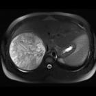
MRI
The tumor itself has the following signal characteristics :
- T1: hypointense
- T2: hyperintense
- T1 C+ (Gd): heterogeneous enhancement
Additionally, there may be focal areas within the tumor of hyperintensity on T1-weighted images and hypointensity on T2-weighted images, which correlate with regions of hemorrhage .
The surrounding pseudocapsule has the following signal characteristics :
- T1: hypointense
- T2: hypointense
Treatment and prognosis
Treatment consists of complete tumor resection with a chemotherapy regimen .
Although prognosis was once quite poor, with the aid of new multimodal treatment the survival rates are improving, with most of the cases currently considered curable .
History and etymology
The term "undifferentiated embryonal sarcoma" was first proposed and used by J Thomas Stocker and Kamal G Ishak, American physicians, in 1978 .
Differential diagnosis
- hepatic mesenchymal hamartoma (younger patients)
- hydatid cyst (in endemic areas)
- hepatic abscess (clinical evaluation)
- cystic degeneration in hepatoblastoma or hepatocellular carcinoma (rare; elevated AFP)
- cystic metastases (rare in children)
See also
Siehe auch:
und weiter:


 Assoziationen und Differentialdiagnosen zu undifferenziertes embryonales Sarkom der Leber: