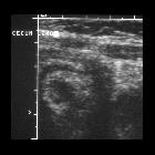
eosinophile Enterokolitis

Eosinophilic
enterocolitis: a case report. MR enterography showing a discontinuous an inflammatory thickening of the ileum (Blue arrows indicate an inflammatory thickening of the ileum)

A rare case
of eosinophilic gastrointestinal disorders with short bowel syndrome after strangulated bowel obstruction. Findings prior to prednisolone administration. a Colonoscopy findings: diffuse redness and edema of the colonic mucosa. b Pathological findings of biopsy from colonoscopy: moderate inflammatory cell infiltration and numerous eosinophil counts, > 20 cells/HPF
eosinophile Enterokolitis
eosinophile Gastroenteritis Radiopaedia • CC-by-nc-sa 3.0 • de
Eosinophilic gastroenteritis (EG) is an uncommon disease characterized by diffuse infiltration of any or all layers of gut wall by eosinophils.
Epidemiology
Eosinophilic gastroenteritis is an uncommon but not rare disease with slight male predominance. It can affect any age group but usually patients present in their 3to 5 decades of life.
Clinical presentation
- self-limiting disorder in the majority of cases
- patients may present with dysphagia, food impaction, abdominal pain, nausea, vomiting, or diarrhea depending on the site of involvement
- in severely affected children it may result in growth retardation
- ascites and pleural effusions may be present
Pathology
- chronic inflammatory disorder of GI wall of undetermined pathophysiology, although atopy-related genes, inflammatory cells, and mediators play a role in the pathogenesis of eosinophilic gastroenteritis
- history of allergy, in particular food allergy, is present in approximately 50% of cases
- more than 60% of cases show peripheral eosinophilia
- on biopsy, there is evidence of mucosal edema and infiltration of the eosinophils, polymorphonuclear leukocytes (PMNs), and lymphocytes in different layers of the gut wall
Location
It can affect various segments of the bowel which include
- esophagus: eosinophilic esophagitis
- stomach: eosinophilic gastritis
- small bowel: eosinophilic enteritis
- large bowel: eosinophilic colitis
Radiographic features
Fluoroscopy
- eosinophilic esophagitis may show a "ringed esophagus" (series of ring-like strictures), and/or smooth long segment narrowing of the esophagus
- gastrointestinal involvement is more common
- virtually always involves gastric antrum and proximal small intestine
- non-specific mucosal fold thickening and nodularity particularly in the gastric antrum, without submucosal edema
- when chronic, the antrum is narrowed with a nodular "cobblestone" mucosal appearance
CT
- CT features are non-specific and may show GI wall thickening and submucosal edema
Treatment and prognosis
- responds well to oral corticosteroids and dietary exclusion
Siehe auch:

 Assoziationen und Differentialdiagnosen zu eosinophile Enterokolitis:


